Lab 27: Dissection: Infratemporal Fossa and Floor of Mouth; Bony Anatomy of Pterygopalatine Fossa
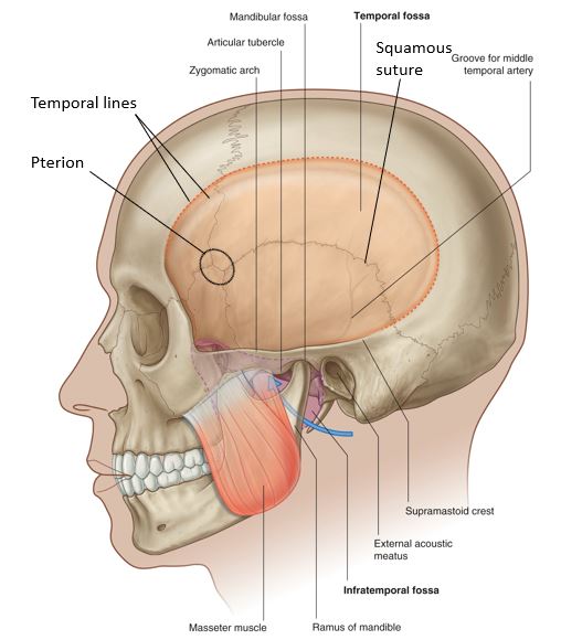
Download this lab as a PDF Goals Identify the temporal fossa and temporalis muscle. On a skull, identify the borders of the infratemporal fossa (ITF) and the foramina that connect it to other regions in the head. Clean the masseter muscle on one side of the face and remove it from the mandible. Use an […]
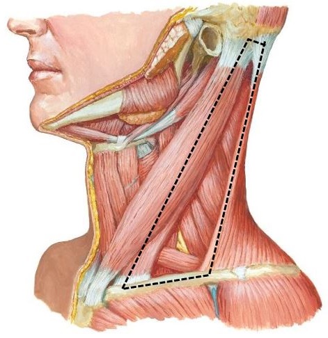
Lab 26: Neck: Anterior Triangle of Neck
Download this lab as a PDF Goals Review the bony anatomy of the cervical vertebrae and the hyoid bone. Clean the muscles and landmarks that define the triangles of the neck. Dissect the contents of the anterior triangle focusing on these subtriangles: carotid, muscular, and submental. Study prosected specimens. Osteology of the Neck Cervical Vertebrae […]
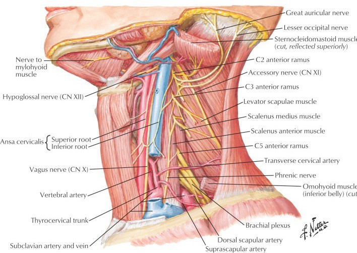
Lab 25: Dissection: Posterior Triangle of Neck, Deep Neck, and Root of Neck
Download this lab as a PDF Goals Clean and identify the contents of the posterior triangle of the neck. Perform a deep dissection to examine the sympathetic trunk and prevertebral muscles. Review the bony anatomy of the superior thoracic aperture and identify its contents. Clean and identify the structures in the root of the neck. […]
Protected: Lab 24: Dissection: Orbit and Ear
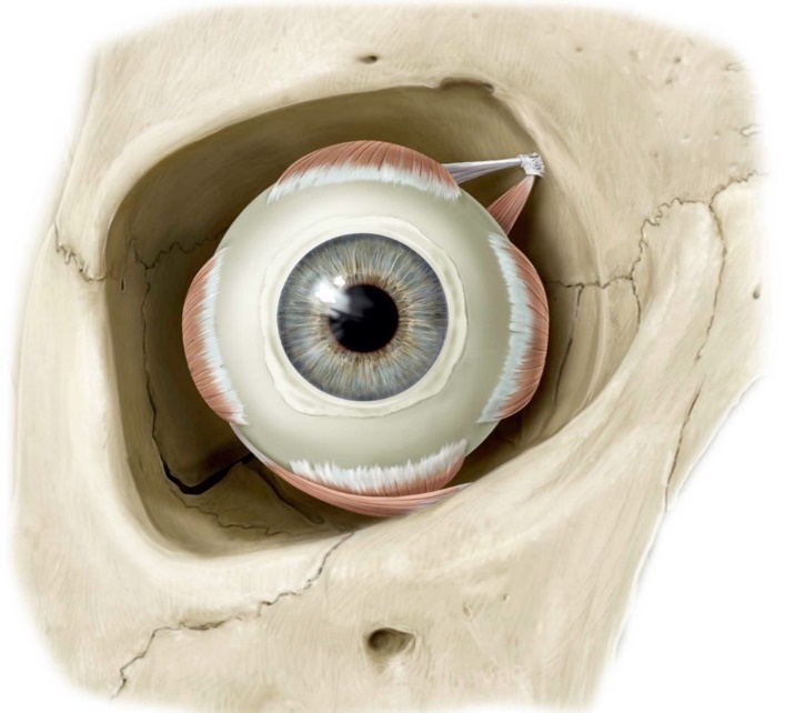
There is no excerpt because this is a protected post.
Protected: Lab 23: Face, Parotid Gland, and Superficial Neck
There is no excerpt because this is a protected post.
Protected: Lab 22: Scalp, Cranial Cavity, and Meninges
There is no excerpt because this is a protected post.
Lab 21, Station 5: Lungs, pleura, trachea, and bronchi
Lab 21 navigation External Anatomy of the Lungs Apex and base Lobes Left lung = superior (upper) and inferior (lower) Right lung = superior (upper), middle, and inferior (lower) Surfaces = costal, mediastinal, and diaphragmatic Anterior and inferior borders In left lung: Cardiac notch and lingula Oblique fissure in both lungs; horizontal fissure in right lung […]
Protected: Lab 21, Station 4: The larynx
There is no excerpt because this is a protected post.
Protected: Lab 21, Station 3: The pharynx
There is no excerpt because this is a protected post.
Protected: Lab 21, Station 2: Skull bones and paranasal sinuses
There is no excerpt because this is a protected post.