old-Lab 24: Peritoneal Cavity and Supracolic Region
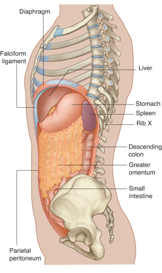
Download this lab as a PDF Goals Survey the GI organs. Examine the peritoneum, peritoneal cavity, mesenteries, omenta, and peritoneal ligaments. Locate the subparts of the greater sac. Study the lesser omentum and demonstrate the lesser sac. Dissect the celiac trunk and demonstrate its branches. Clean and study the organs in the supracolic region: liver; […]
Protected: Lab 7: Posterior Forearm and Dorsal Hand
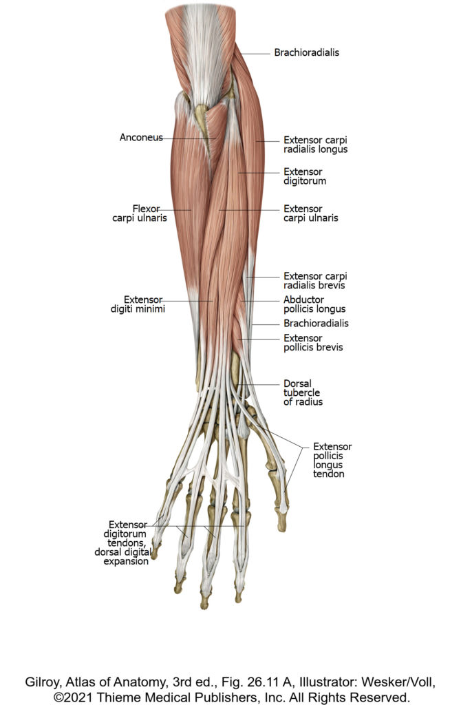
There is no excerpt because this is a protected post.
Protected: Lab 22, Station 1: Pelvic Skeleton and Ligaments
There is no excerpt because this is a protected post.
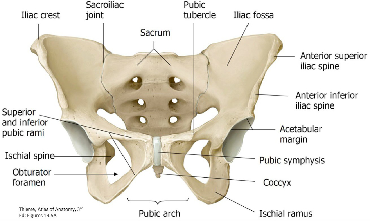
Lab 12, Station 1: Pelvic Skeleton and Ligaments
Lab 12 Navigation Introduction Station 1: Pelvic Skeleton and Ligaments Station 2: Pelvic Cavity and Anal Triangle of Perineum Station 3: Female Perineum (Urogenital Triangle) Station 4: Male Perineum (Urogenital Triangle) Station 5: Overview: Pelvic Organs and Vessels Station 1: Pelvic Skeleton and Ligaments Complete anatomy Hip bone Pelvic Skeleton (Pelvic Girdle): Two Hip Bones […]
Protected: Lab 22, Station 2: Pelvic Cavity and Anal Triangle of Perineum
There is no excerpt because this is a protected post.
Lab 22: Prosection lab: Pelvic Skeleton, Pelvic Cavity, and Pelvic Vessels; Perineum
Students: Unlike most lab sessions, which are dissection-based, this session is a “prosection lab.” Assemble in 5 groups, each having ~16 members. There are 5 learning stations situated around the labs. Pick a station to start at. Groups will spend about 25 minutes at each station, rotating around the labs until they have visited all […]
Protected: Lab 22, Station 5: Overview: Pelvic Organs and Vessels
There is no excerpt because this is a protected post.
Protected: Lab 22, Station 4: Male Perineum (Urogenital Triangle)
There is no excerpt because this is a protected post.
Protected: Lab 22, Station 3: Female Perineum (Urogenital Triangle)
There is no excerpt because this is a protected post.
Protected: Lab 6: Carpal Tunnel and Palmar Hand
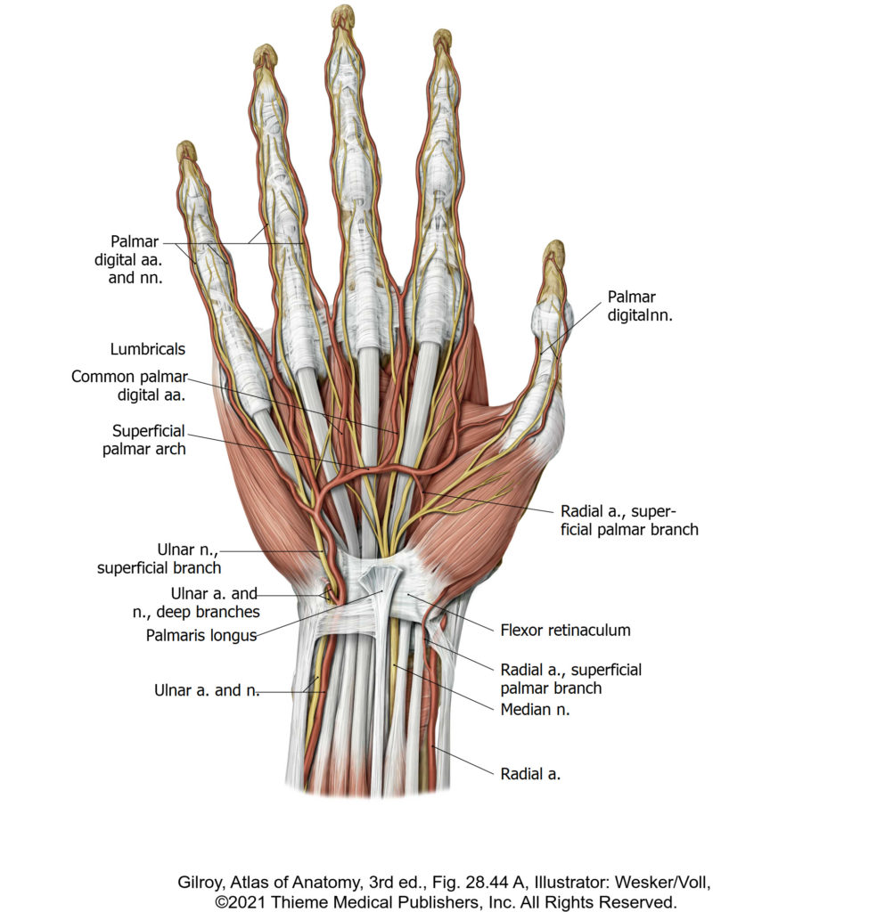
There is no excerpt because this is a protected post.