

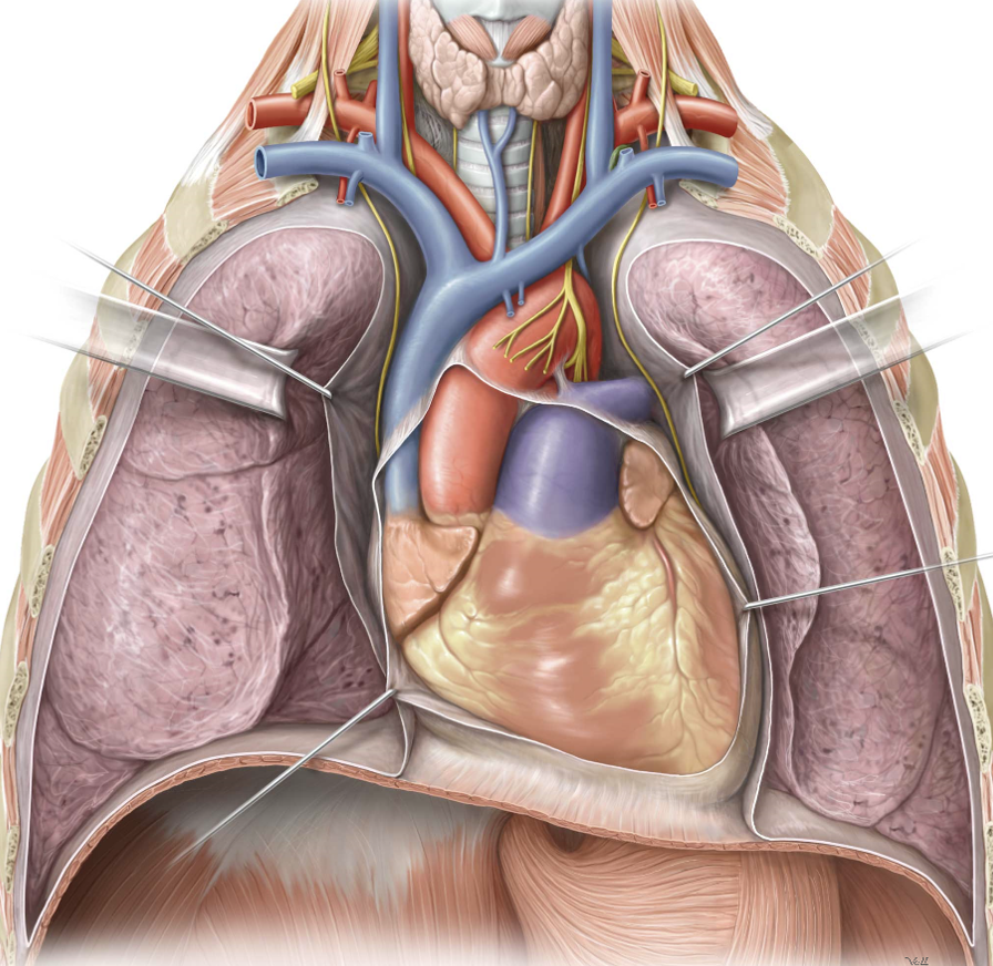
Thoracic cavity
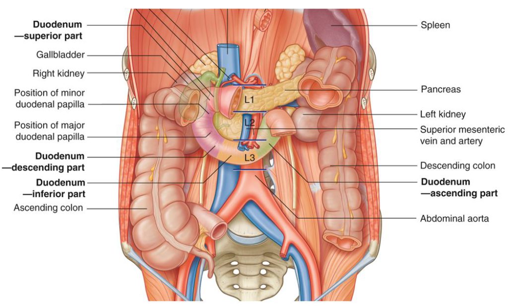
Protected: Lab 25: Infracolic Region
There is no excerpt because this is a protected post.
Protected: Lab 23: Oral Cavity, Pharynx, and Muscles of Mastication
There is no excerpt because this is a protected post.
Protected: Lab 23, Station 5: Oral Region and Salivary Glands—Lateral View
There is no excerpt because this is a protected post.
Protected: Lab 23, Station 4: Oral Cavity and Pharynx—Sagittal View
There is no excerpt because this is a protected post.
Lab 23, Station 3: Nerves and Vessels Associated with the Oral Cavity
Lab 23 navigation Station 3: Nerves and Vessels Associated with the Oral Cavity Branches of V3 in the Infratemporal Fossa Mandibular nerve (V3). Enters the infratemporal fossa via foramen ________________ Its branches include: Inferior alveolar nerve: Supplies lower teeth Lingual nerve: Supplies the anterior 2/3 of tongue with general sensation Chorda tympani nerve (from CN […]
Protected: Lab 23, Station 2: Muscles of Mastication and TMJ
There is no excerpt because this is a protected post.
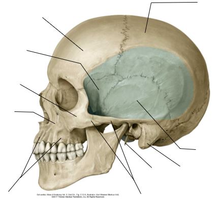
Lab 23, Station 1: Osteology Associated with the Oral Cavity
Lab 23 navigation Station 1: Osteology Associated with the Oral Cavity This station will be self-guided. Use the skull bones and laminated images provided to practice identifying the structures listed. Frontal bone Parietal bone Temporal bone Zygomatic bone Maxilla Body (contains maxillary air sinus) Alveolar process (contains tooth sockets) Figure 23.1. (Drag the slider to […]
Lab 11: Peritoneal Cavity and Supracolic Region
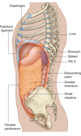
Download this lab as a PDF Goals Survey the GI organs. Examine the peritoneum, peritoneal cavity, mesenteries, omenta, and peritoneal ligaments. Locate the subparts of the greater sac. Study the lesser omentum and demonstrate the lesser sac. Dissect the celiac trunk and demonstrate its branches. Clean and study the organs in the supracolic region: liver; […]
Lab 10: Dissection: Anterior Abdominal Wall (AAW) and Inguinal Region
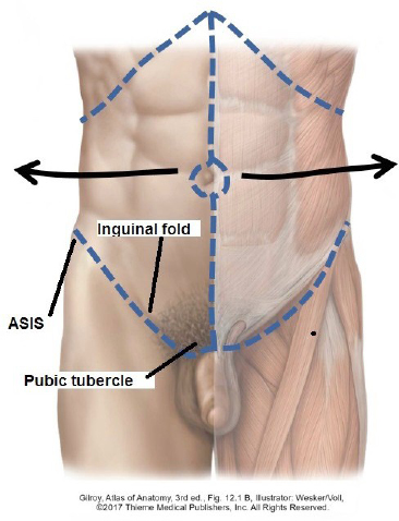
Download this lab as a PDF Goals Reflect the skin from the anterior and lateral abdominal wall. Identify the two layers of the superficial fascia and reflect them. Clean and identify the external oblique, internal oblique, and transversus abdominis muscles. Identify and open the rectus sheath on one side and identify the rectus abdominis muscle. […]