Lab 28: Neck: Anterior Triangle of Neck

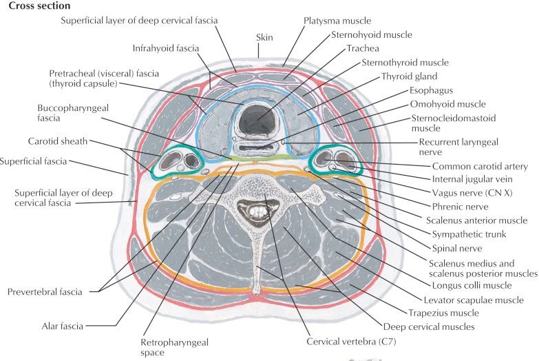
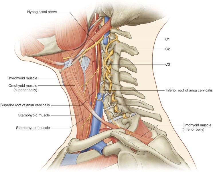
1Review the bony anatomy of the cervical vertebrae and the hyoid bone.

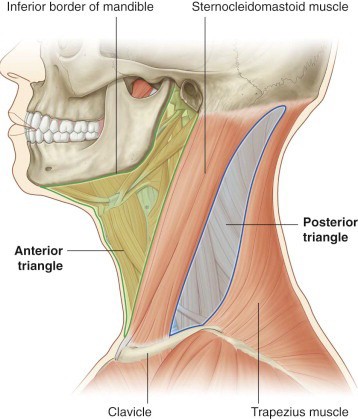
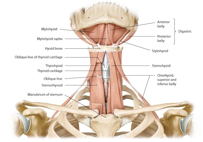
2Clean the muscles and landmarks that define the triangles of the neck.

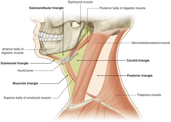
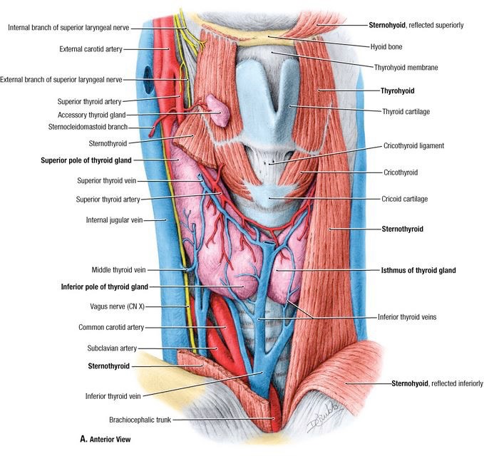
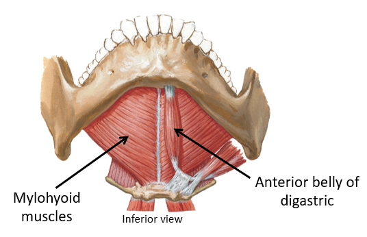
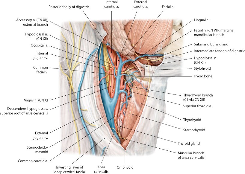
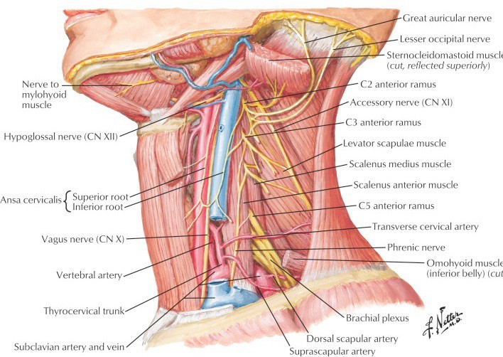
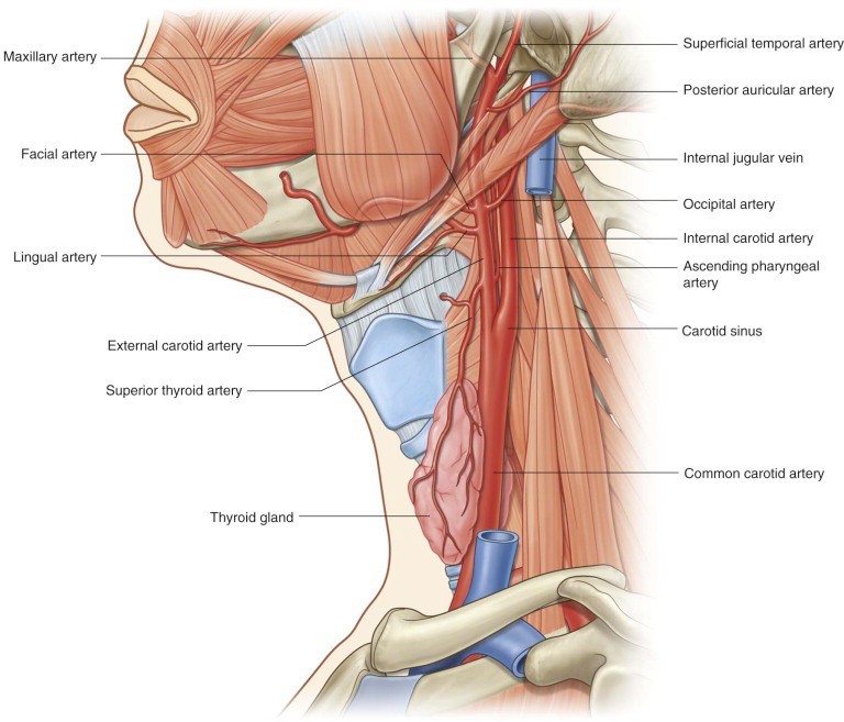
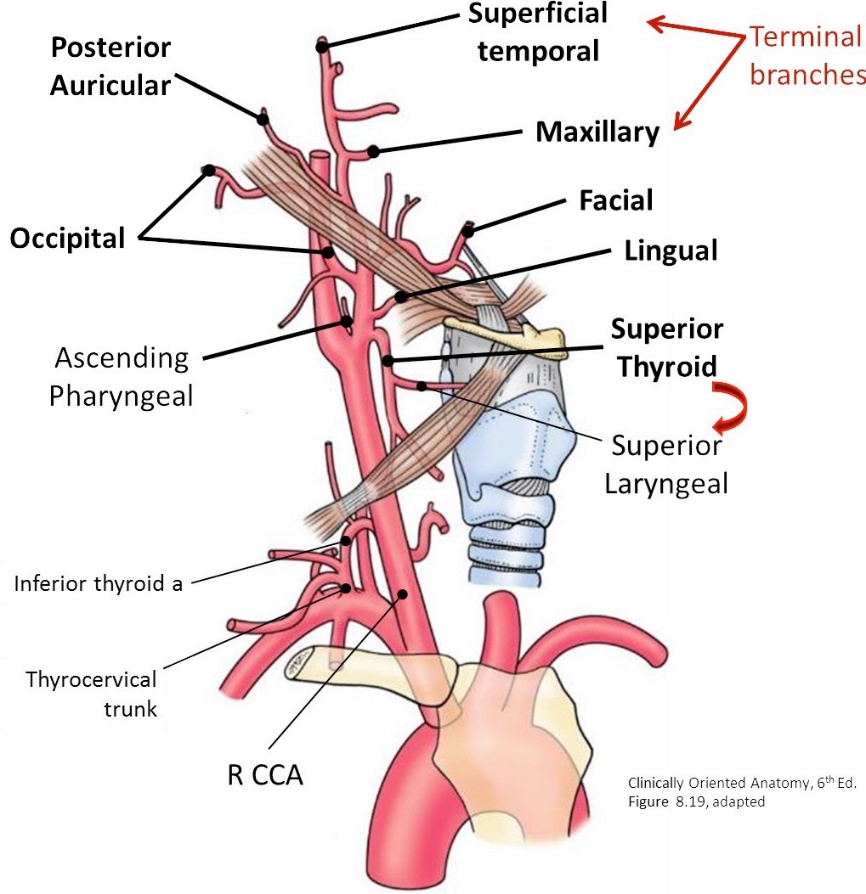
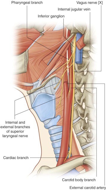
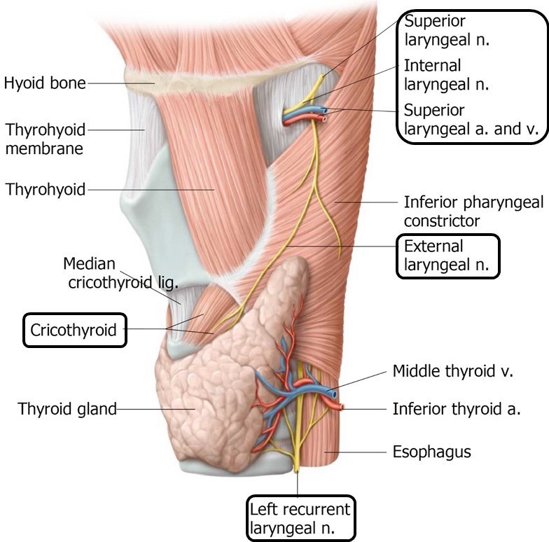
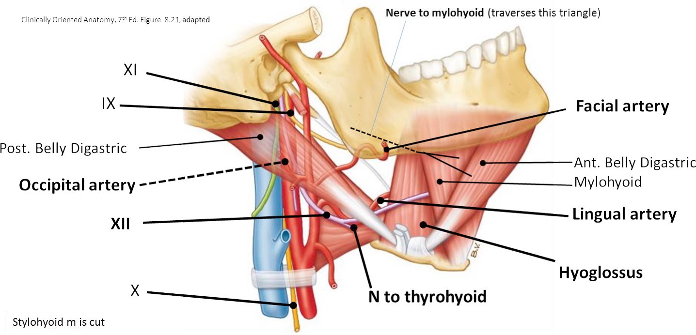
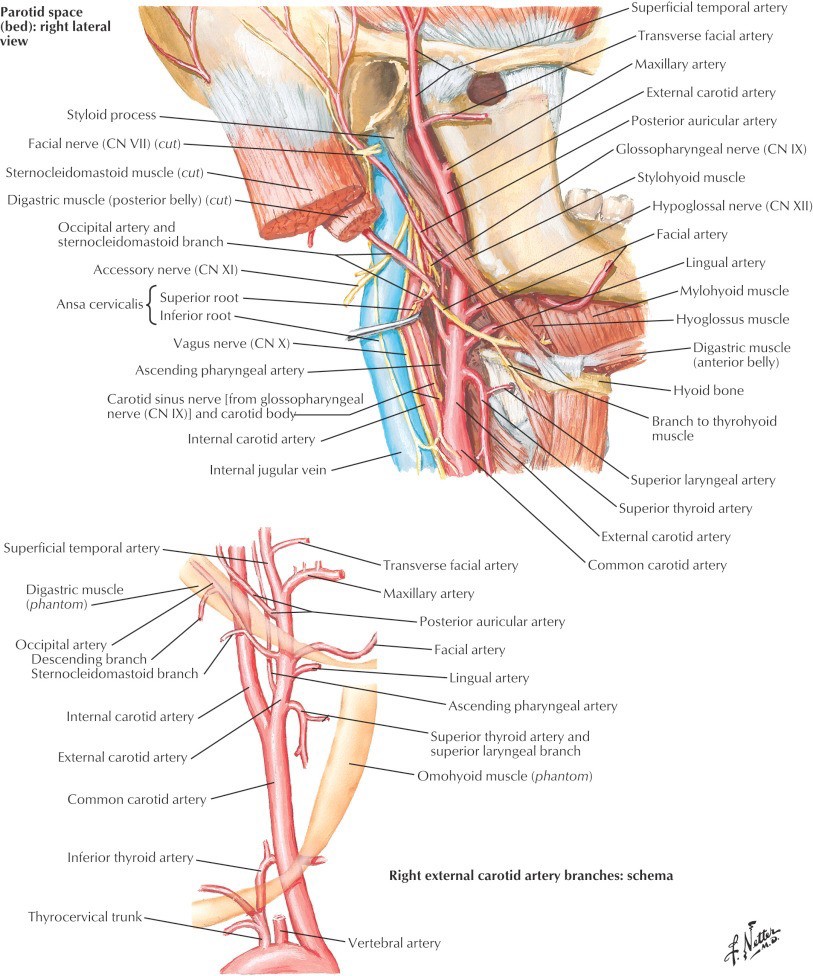
3Dissect the contents of the anterior triangle focusing on these subtriangles: carotid, muscular, and submental.

4Study prosected specimens.


Cervical Vertebrae and Hyoid Bone

Atlas (C-1)

Lateral masses—with superior and inferior articular surfaces

Anterior and posterior arches

Axis (C-2)

Body

Dens

Vertebral arch (with pedicles and laminae)

C-3 through C-7

Body

Vertebral arch

Pedicle

Lamina

■•Note that the spinous processes are bifid.

General Features of Cervical Vertebrae

All cervical vertebrae have a transverse foramen in their transverse processes. The vertebral arteries pass through the foramina of C-1 to C-6 but miss the foramina of C-7.

Note the intervertebral foramina formed when two adjacent vertebrae articulate these transmit cervical spinal nerves above their corresponding vertebrae (i.e., C-2 spinal nerve exits above C-2 vertebra).

Between which bones does the C-1 spinal nerve exit?

Hyoid Bone

Body of hyoid bone

Greater and lesser horns

The hyoid is in the anterior neck at the level of C-3.

The hyoid is unique in that does not articulate with any other bone.

Supra-hyoid muscles attach the hyoid to the mandible and styloid processes.

Infra-hyoid muscles (strap muscles) attach the hyoid to the thyroid cartilage, sternum, and scapulae.

Cervical Fascia

In a previous lab (face and superficial neck), the skin was reflected from the neck.

In the superficial fascia of the neck identify the following:

Platysma muscle

External jugular veins

Anterior jugular veins

Reflect the platysma muscles.

Reflect the platysma muscles superiorly toward the face. Grasp the thin muscle with forceps and carefully peel it away from the subcutaneous tissue, using a scalpel or scissors. Leave the remaining superficial fascia in the neck intact.

Figure 28.1.

Deep to the superficial fascia of the neck is the deep cervical fascia. It has these subparts:

Investing fascia

Pretracheal fascia

Prevertebral fascia

Carotid sheath

Chalk Talk

Draw a transverse section of the neck, and sketch in the three layers of deep cervical fascia: investing, pretracheal, and prevertebral. Include the infrahyoid part of the pretracheal fascia = it encloses the infrahyoid (strap) muscles. Also draw in the carotid sheath.

Figure 28.2.

The investing fascia forms the roof of the triangles of the neck. It forms envelops around the sternocleidomastoid and trapezius muscles. It must be incised and cleaned in order to view the contents of the neck triangles.

Triangles of the Neck

Figure 28.3.
Figure 28.4.

Chalk Talk

Sketch and identify the boundaries of the anterior and posterior triangles. Then sketch and describe the boundaries of subtriangles in the neck: submandibular, submental, carotid, and muscular triangles.

Clean and reflect the sternocleidomastoid muscles.

Carefully clean the investing fascia from the surface of the sternocleidomastoid muscles and reflect the muscle on both sides superiorly, elevating the muscles toward the head.

Anterior Triangle

Locate and palpate the following:

Landmarks

Hyoid bone

Thyroid cartilage of larynx w/ laryngeal prominence (“Adam’s apple”)

Cricoid cartilage of larynx

Cricothyroid membrane

Trachea

Figure 28.5.

Submental Triangle

Not much to see here, folks. Use the anterior bellies of the digastric muscles to define the boundaries of this unpaired triangle that straddles the midline of the neck. The mylohyoid muscle forms the floor of the triangle.

Figure 28.6.

Carotid Triangle

Figure 28.7.

Define the boundaries of the carotid triangle.

Identify the carotid sheath within the carotid triangle and LIST ITS CONTENTS.

The carotid sheath is continuous with all three layers of the deep cervical fascia = investing, pretracheal, and prevertebral. Deep to the sheath is the sympathetic trunk (next lab).

Carefully probe and dissect the surface of the carotid sheaths.

Look for the ansa cervicalis, a motor nerve of the cervical plexus, shaped like a “loop” (superior root from C-1 and an inferior root from C-2,3).

Start out by probing just lateral to the internal jugular vein at the level of the carotid bifurcation to find the inferior root—gently pull it away from the vein and follow it inferiorly as it loops.

Figure 28.8.
Open the carotid sheath with scissors, but try and keep the ansa cervicalis.

Identify:

Internal jugular vein

Common carotid, internal carotid, and external carotid arteries

Vagus nerve

Figure 28.9.

See if you can identify some deep cervical nodes (often on the surface of the vein) located near the omohyoid and digastric muscles.

One of the most prominent of the deep cervical nodes is located superiorly, adjacent to the angle of the mandible. This is the jugulo-digastric node, also known as the “tonsillar” node.

Note the complexity and variability of veins that drain into the internal jugular. Locate the common facial vein.

Figure 28.10.

Follow the internal jugular vein inferiorly to where it joins the subclavian vein to form the brachiocephalic vein. The junction of these veins is often referred to as the jugulosubclavian angle or simply the “venous angle”.

Figure 28.11.

Carotid Arteries

Locate the bifurcation of the common carotid artery at the level of the upper border of the thyroid cartilage (larynx)—about C-4.

The somewhat distended proximal part of the internal carotid artery is the carotid sinus.

Figure 28.12.

What is the function of the carotid sinus? Which nerve carries visceral afferent signals from it?

Within the bifurcation (between ICA and ECA) is a small vascular structure = no bigger than a pin’s head. This is the carotid body.

What is the function of the carotid body? Which nerve carries visceral afferent signals from it?

Clean the posterior belly of the digastric and stylohyoid muscles.

Use the hyoid bone as a landmark to identify the tendon between the anterior and posterior bellies of the digastric muscles. Clean and trace the posterior digastric towards its attachment to the mastoid process. 

The stylohyoid muscle runs from the styloid process to the hyoid bone. Note that the stylohyoid muscle splits around the tendon of the digastric muscle to attach to the hyoid bone. 

What nerve innervates the posterior digastric and stylohyoid muscles? (Hint: the nerve exits the foramen located between the proximal attachment of these two muscles)

Clean the external carotid artery and its branches.

IDENTIFY as many branches of the external carotid in the neck as possible:

From the anterior side of the artery:

Superior thyroid

Lingual

Facial

From the medial side of the artery:

Ascending pharyngeal

From the posterior side of the artery:

Occipital

Posterior auricular

Terminal branches

Maxillary

Superficial temporal

Figure 28.13.
ONE side: reflect the stylohyoid and posterior digastric muscles.

On one side of the neck (preferable, on the same side as the deep dissection of the face where the parotid gland was removed), detach the muscles from the hyoid bone and reflect them. Now trace the external carotid artery up to the head to where the parotid gland used to be located.

Chalk Talk

Sketch the external carotid artery and its branches: three anterior branches, one medial branch, two posterior branches, and two terminal branches. Discuss the regions of the head and neck that receive blood supply from these arteries.

Vagus Nerves

Identify the vagus nerve by separating the common carotid artery and internal jugular vein at the level of the thyroid cartilage. Trace it inferiorly into the thorax.

Find the large superior laryngeal branch of the vagus. This comes off the vagus high, just below the skull. You won’t find the origin of this nerve, but you will see it heading toward the larynx.

The superior laryngeal nerve divides into a thin external branch (external laryngeal nerve) and a larger internal branch (internal laryngeal nerve).

Figure 28.14.

The internal laryngeal nerve enters the larynx through the thyrohyoid membrane. Palpate the hyoid bone then move inferiorly to feel the soft thyrohyoid membrane. The superior laryngeal artery (a branch of the superior thyroid artery) accompanies the internal laryngeal nerve into the larynx.

Relationship: The external laryngeal nerve courses in tandem with the superior thyroid artery. Earn a gold star for the day by finding this thin nerve. It courses inferiorly to the cricothyroid muscle of the larynx.

Figure 28.15.

What is the function of the internal and external laryngeal nerves?

(Hint: Think back to the respiratory block when you learned about the larynx; one branch is sensory and one is motor.)

Submandibular Triangle

Figure 28.16.

Identify the boundaries of the submandibular triangle.

Why is this also known as the digastric triangle?

Identify the submandibular gland, anterior and posterior bellies of the digastric muscle, stylohyoid muscle, and facial artery and vein. The facial artery is very tortuous as it passes around the submandibular gland and over the mandible to the face.

Note any submandibular lymph nodes.

Figure 28.17.

Locate and clean the hypoglossal nerve (CN XII).

Tip

Locate the posterior belly of the digastric and occipital artery as landmarks.

The lowest part of the nerve loops below the posterior belly of the digastric/stylohyoid muscles.

In almost every case, the hypoglossal nerve crosses the occipital artery—so look for this sure-fire relationship.

The hypoglossal continues above the hyoid bone into the base of the tongue.

What is the function of CN XII?

Nerve fibers from the ventral ramus of C-1 “hitch a ride” on the hypoglossal to get where they need to go = they drop off to form the superior root of the ansa cervicalis, as well as to innervate the thyrohyoid and geniohyoid muscles.

Finally, clean the deep surface of the reflected sternocleidomastoid muscles (SCM). Look for the flat, white, thick spinal accessory nerve entering the deep surface of the SCM.

Muscular Triangle

It contains the infrahyoid (strap) muscles.

Clean the sternohyoid muscles and superior bellies of the omohyoid. The fascia you are removing from the infrahyoid muscles is called the infrahyoid layer of the pretracheal fascia.

Figure 28.18.
One side: Reflect the sternohyoid muscle on one side of the neck.

1Identify the sternothyroid and thyrohyoid muscles deep to the reflected sternohyoid.

2Now reflect the sternothyroid muscle to see the thyroid gland under it. The thryoid is surrounded by a capsule made of pretracheal fascia.

3 Locate the thyroid cartilage of the larynx, the cricoid cartilage, and the cricothyroid membrane. The cricothyroid membrane is the “soft spot” between the thyroid and cricoid cartilages of the larynx.

Clinical correlation

The cricothyroid membrane is the site of an “emergency airway” (cricothyrotomy).

In emergent situations (CICO = “cannot intubate, cannot oxygenate”), an emergency airway can be created via the cricothyroid membrane. Cricothyrotomy is performed by inserting a tube through an incision in the skin and cricothyroid membrane. This site is preferred because it is superficial, palpable, and usually without anatomic obstructions.

Checklist, Lab #28

Review your dissection and make sure you have identified each of the structures in the checklists that follow.

Note

Checklists for the cervical vertebrae and the hyoid bone are in the Osteology section of this lab guide.

Review boundaries of neck triangles and landmarks in the anterior neck

Anterior and posterior triangles

Submental, submandibular, carotid, and muscular triangles

Hyoid bone

Thyroid cartilage w/laryngeal prominence

Cricoid cartilage

Cricothyroid membrane

Trachea

Divisions of deep cervical fascia

Investing

Prevertebral

Pretracheal

Infrahyoid part of pretracheal fascia

Carotid sheath

Muscles

Sternocleidomastoid

Trapezius

Digastric (anterior and posterior bellies)

Stylohyoid

Mylohyoid

Omohyoid (superior and inferior bellies)

Sternohyoid

Sternothyroid

Thyrohyoid

Nerves

Ansa cervicalis

Phrenic nerve

Spinal accessory nerve (entering the deep surface of the SCM)

Hypoglossal nerve

Vagus

Superior laryngeal nerve (branch of vagus)—find the internal branch (internal laryngeal nerve) entering the larynx through the thyrohyoid membrane just below the hyoid bone

Vessels

Internal jugular vein

Subclavian artery and vein

Common carotid artery

Internal cartoid artery

External carotid artery

Carotid sinus and carotid body

Superior thyroid artery

Facial artery

Lingual artery

Occipital artery (wish list item—follow the hypoglossal nerve proximally—it crosses deep to the occipital artery)

Other

Deep cervical lymph nodes

Jugulodigastric node (a specially named deep cervical node)—the jugulodigastric node is the “tonsillar node.” Why?

Table of Contents