Five critical electrical and mechanical functions.
Five parts of the heart’s electrical system.
SA node.
P wave.
AV node.
Bundle of His.
Right and left bundle branches.
Purkinje fibers.
QRS complex.
The heart has an intricate electrical system, made up of highly specialized cells, that is responsible for generating each heart beat. The specialized cells are responsible for five critical electrical and mechanical functions:
Establishing the ability to create an automatic and regular heart rhythm.
Allowing communication among the hundreds of millions of cells in less than one-tenth or two-tenths of a second.
Providing for activation of the myocardial cells.
Triggering mechanical contraction.
Resetting the system for the next cycle in a process called repolarization. Except for mechanical contraction, all of these functions are represented on the EKG.
The heart’s electrical system
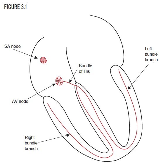
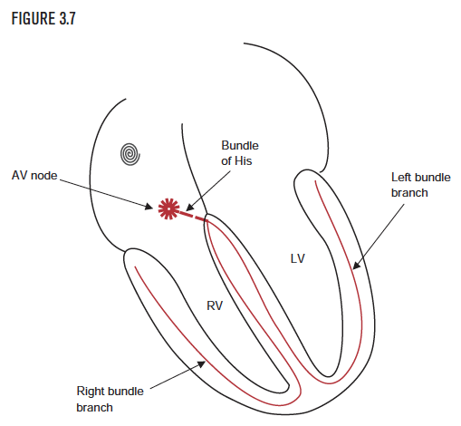
The heart’s electrical system consists of five structures:
The sinoatrial (or SA) node.
The atrioventricular (or AV) node.
The bundle of His.
The right and left bundle branches.
The Purkinje fibers.
The right and left atria contract in atrial systole, as the electrical impulse triggers atrial contraction. The right and left ventricles contract in ventricular systole, as the electrical impulse triggers mechanical contraction (Figure 3.1).
Creation of the rhythm:
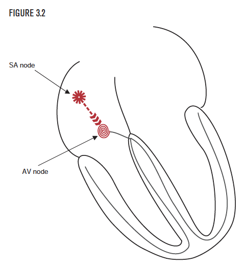
The sinus node
The sinoatrial node (SA node) is the heart’s natural pacemaker because it normally initiates each heartbeat and maintains the rest of the heart’s pace. The SA node comprises hundreds of specialized cells and is located in the upper part of the right atrium. The SA node normally generates an average of 60 to 100 impulses per minute. These impulses that travel through the atria via the internodal conduction pathway cause the atria to contract. This depolarizes both atria, as the signal also heads for the AV node (Figure 3.2). This electrical activation of the atria (called depolarization) is represented on the EKG as a “P” wave. This is normally the first wave, or “deflection,” on the EKG.
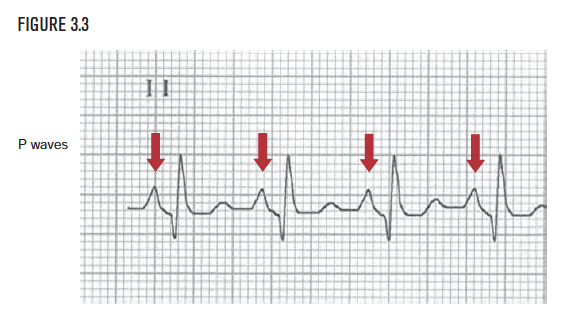
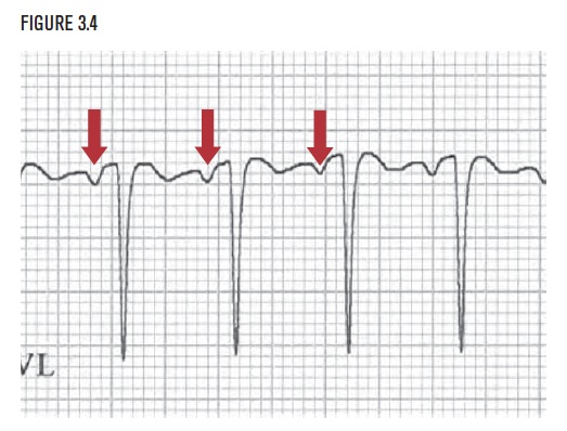
Identifying the P wave on the EKG
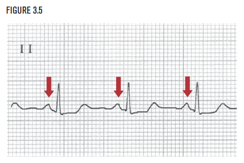
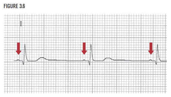
Since the normal orderly process begins when the SA node depolarizes the right and left atria, we look for and see the P wave first. The size, shape, and amplitude of the P wave may vary. It may occasionally be small and difficult to find. The P wave may be mostly below the baseline (as in Figure 3.4), mostly above the baseline (as in Figure 3.5 below), or mixed. The P wave may be relatively tiny and difficult to see (as in Figure 3.6). The apparent absence of a P wave is abnormal and suggests that the rhythm may be originating from an ectopic pacemaker in the atrial wall, or in the atrioventricular junction. (See Section III for more on this subject.)
Communication with the ventricles:
The atrioventricular node and the bundle branches
The atrioventricular node (AV node) is located in the lower part of the right atrium. The AV node receives the impulse from the SA node and continues transmitting (communicating) it to the bundle of His.
The bundle of His is found below the AV node and communicates (conducts) the electrical impulse through to the bundle branches. The bundle branches divide into the right bundle branch, which leads to the right ventricle, and the left bundle branch, which leads to the left ventricle. The electrical stimulus travels down the bundle branches to the Purkinje fibers. Finally the myocardial cells contract, a mechanical event that is known as ventricular systole. For a detailed discussion of normal and abnormal conduction, see Chapters 9, Chapter 10, Chapter 11, and Chapter 12.
Depolarization of the ventricles:
The QRS complex
Once the impulse reaches the Purkinje network, it spreads (communicates) onward and activates, or depolarizes, the myocardial cells. This is referred to as ventricular depolarization. Ventricular depolarization is represented on the EKG as the QRS complex.
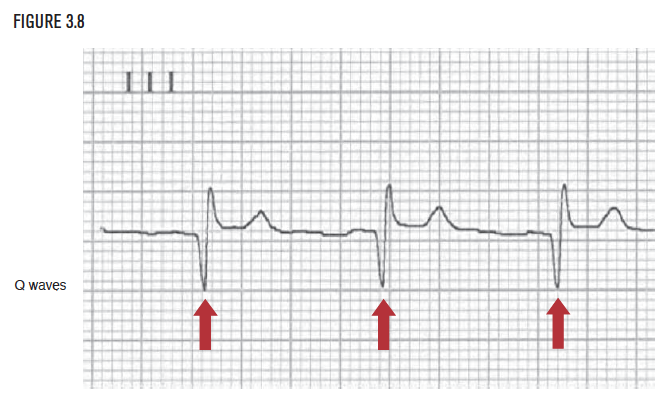
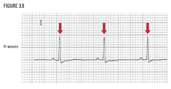
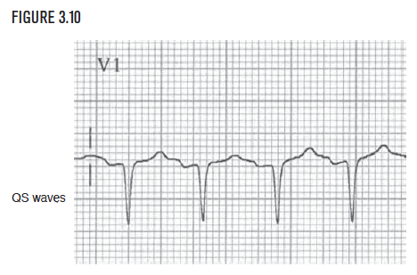
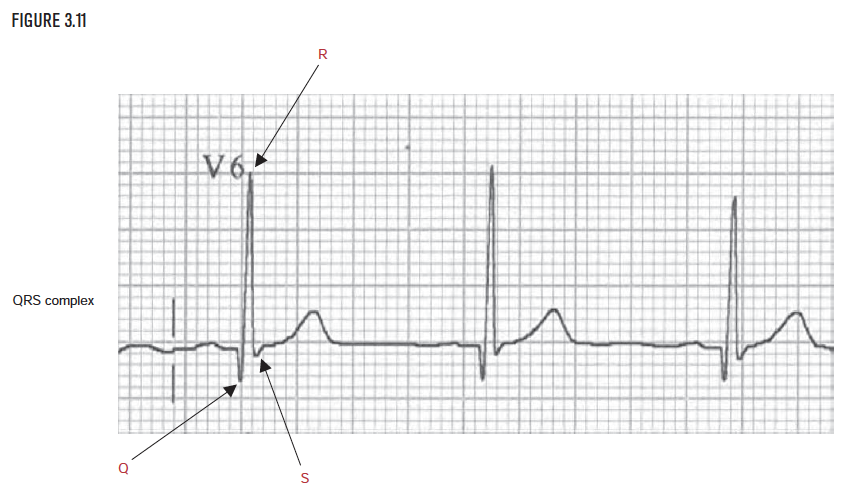
The QRS complex is the second deflection on the normal EKG. The QRS complex consists of a Q wave, an R wave, and/or an S wave, occurring either singly or in any combination. Although the complex is called the QRS complex, it does not always contain a Q wave, an R wave, and an S wave. If the first wave of the complex is a downward deflection, as in Figure 3.8, the complex is considered to have a Q wave. The next upward wave is called an R wave. If the first wave of the QRS is upward, as in Figure 3.9, then this wave is called an R wave. The downward wave that follows is an S wave. Note that although the EKG in Figure 3.9 does not contain a Q wave, the whole complex is still called a QRS! When no R wave is present at all, as in Figure 3.10, these deflections are called QS waves. Finally, a QRS can have all three waves, as in Figure 3.11.
Repolarization of the ventricles:
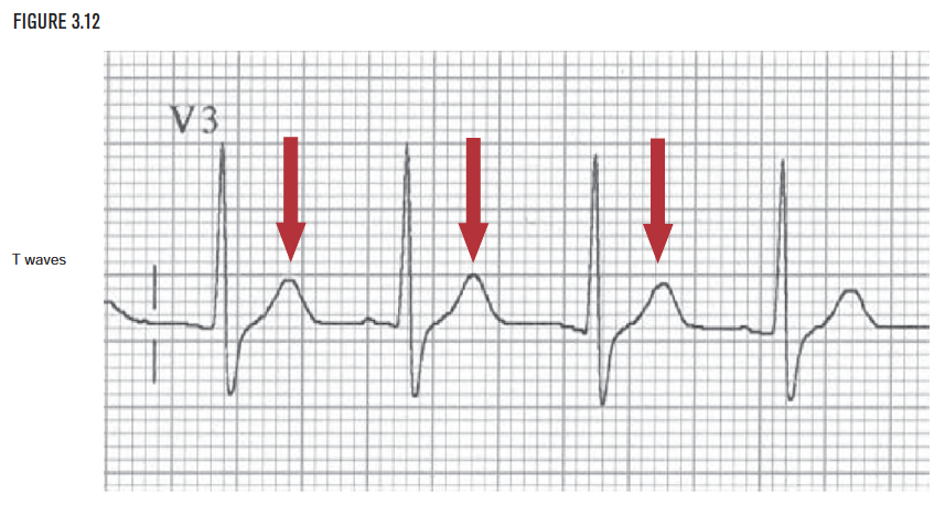
The T wave
The third deflection on the EKG is the T wave. The T wave represents ventricular repolarization. The ventricles must repolarize or recharge themselves before the next cardiac cycle can begin.