Clinical Conditions that Affect the EKG Worksheets

Headshot of Kevin Hodges, Vice Chair, Emergency Medicine
Kevin Hodges
Vice Chair, Emergency Medicine
envelope icon phone icon
Headshot of Chris Anderson, Vice Chair, Pediatrics
Chris Anderson
Vice Chair, Pediatrics
envelope icon

These worksheets are for self-study only. Answers will not be evaluated.

Instructions for Chapter 20 Worksheets

  1. Make basic measurements, evaluate for ischemia, infarction, and hypertrophy.
  2. Diagnose clinical conditions based on criteria described in Chapter 20.
  3. Evaluate clinically.


Clinically-Based Critical Thinking: Interpretation

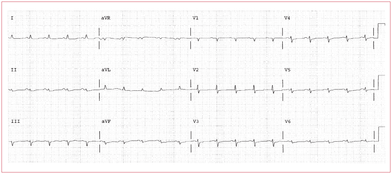
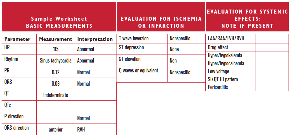
There is sinus tachycardia associated with low voltage. Possibilities include COPD (with sinus tachycardia due to hypoxia or sympathomimetic therapy), or a large pericardial effusion, or tamponade. The presence on an anterior QRS is consistent with pulmonary hypertension. The low amplitude affects the T waves and P waves as well.

Worksheet 20.1

Complete the basic measurements, evaluate for ischemia, infarction, and hypertrophy, diagnose clinical condition, and evaluate clinically.

Parameter Measurement Interpretation
HR    
Rhythm    
PR    
QRS    
QT    
QTc    
P direction    
QRS direction    
Abnormal parameter If present, note the leads or location
Inverted T waves  
ST depression  
ST elevation  
Q waves or equivalents  
Systemic effects If present, note
LAA/RAA/LVH/RVH  
Drug effect  
Hyper/hypokalemia  
Hyper/hypocalcemia  
Low voltage  
SI/QT III pattern  
Pericarditis  

Diffuse wide QRS in sine wave pattern. Hyperkalemia.

Worksheet 20.2

Complete the basic measurements, evaluate for ischemia, infarction, and hypertrophy, diagnose clinical condition, and evaluate clinically.

Parameter Measurement Interpretation
HR    
Rhythm    
PR    
QRS    
QT    
QTc    
P direction    
QRS direction    
Abnormal parameter If present, note the leads or location
Inverted T waves  
ST depression  
ST elevation  
Q waves or equivalents  
Systemic effects If present, note
LAA/RAA/LVH/RVH  
Drug effect  
Hyper/hypokalemia  
Hyper/hypocalcemia  
Low voltage  
SI/QT III pattern  
Pericarditis  

Sinus Tachycardia. RVH. SI QT III pattern. Rule out pulmonary embolism. Diffuse ST segment depression, so First Rule of the T waves also applies.

Worksheet 20.3

Complete the basic measurements, evaluate for ischemia, infarction, and hypertrophy, diagnose clinical condition, and evaluate clinically.

Parameter Measurement Interpretation
HR    
Rhythm    
PR    
QRS    
QT    
QTc    
P direction    
QRS direction    
Abnormal parameter If present, note the leads or location
Inverted T waves  
ST depression  
ST elevation  
Q waves or equivalents  
Systemic effects If present, note
LAA/RAA/LVH/RVH  
Drug effect  
Hyper/hypokalemia  
Hyper/hypocalcemia  
Low voltage  
SI/QT III pattern  
Pericarditis