Pathophysiology
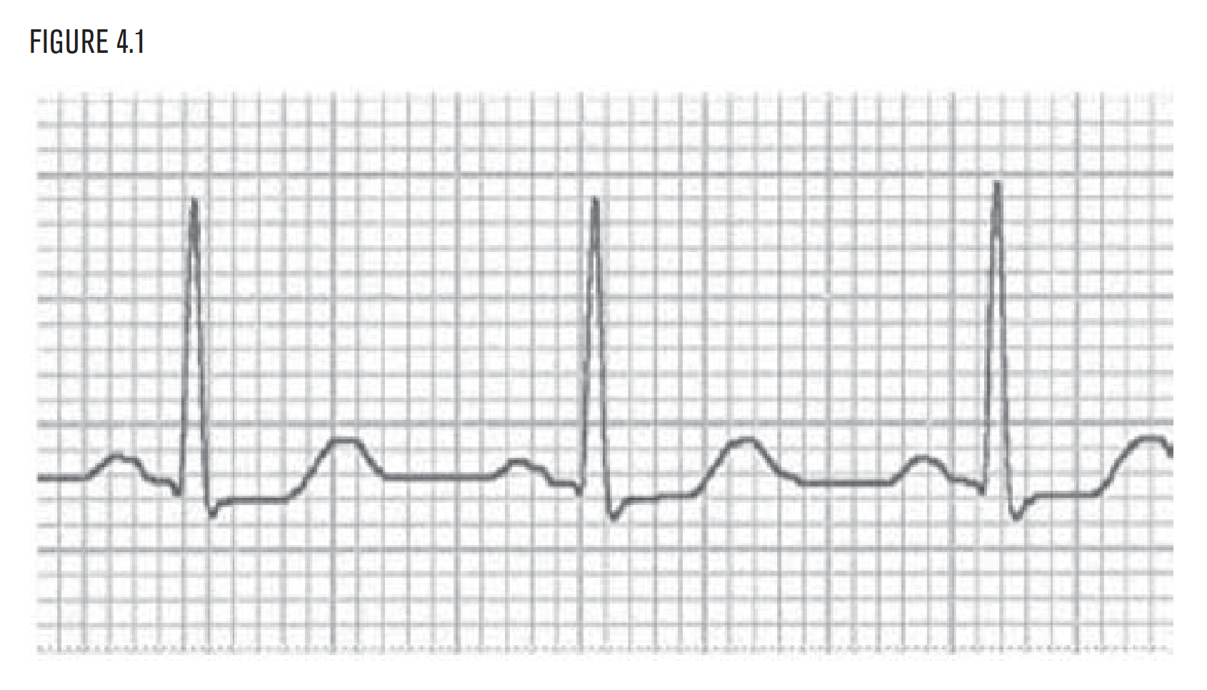
Sinus tachycardia represents a relative imbalance in the normal sympathetic/parasympathetic balance of the heart. There are two basic causes of sinus tachycardia:
- Increased sympathetic activity
- Decreased parasympathetic activity.
Increased sympathetic activity is by far more common clinically. Increased sympathetic activity is part of the ancient “flight or fight” protective emergency system of the body. It is analogous to someone inside the patient’s body calling a “code” or pulling a fire alarm. It is that serious, and that significant. The underlying cause should always be determined. It will typically be very relevant clinically.
Causes of increased sympathetic activity include:
- Shock.
- Heart failure.
- Infection.
- Bleeding.
- Pain.
- Pulmonary embolism.
- Hypoxia.
- Hypoglycemia.
- Anxiety.
- Drugs.
 All are major disorders. Commonly used drugs that cause increased sympathetic activity include bronchodilators, inotropic infusions, and pressors. From the EKG, diagnose sinus tachycardia, and then evaluate the patient to determine its cause. Decreased parasympathetic activity is a much less common cause of sinus tachycardia and is most commonly related to atropine administration or poison ingestion. The most common big mistake EKG readers make is ignoring the presence of sinus tachycardia. A return to the bedside frequently provides the answer!
Pathophysiology
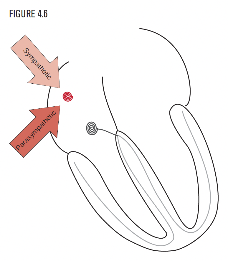
Sinus bradycardia represents a relative imbalance in the normal sympathetic/ parasympathetic balance of the heart. There are two basic causes of sinus bradycardia:
- Decreased sympathetic activity.
- Increased parasympathetic activity.
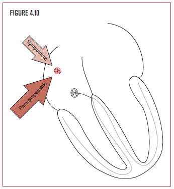 Parasympathetic activity is frequently the “relax and take your time” signal that counterbalances the sympathetic nervous system. Increasing parasympathetic activity slows down the heart rate. Decreased sympathetic activity does the same and is more common clinically. It is the result of the current common practice of using drugs that block the sympathetic nervous system in the treatment of hypertension, coronary artery disease, and heart failure. It is also possible to directly stimulate the parasympathetic nervous system. A common cause of increased parasympathetic activity is the vagal response. The vagal response can occur secondary to gastrointestinal (GI) stimulation during nausea and vomiting, drug treatment, the carotid reflex, or with direct therapeutic vagal stimulation for seizures or depression.