


These worksheets are for self-study only. Answers will not be evaluated.
Worksheet 1
??Examine the whole strip and determine an atrial and ventricular rate??
| Parameter | Measurement | Interpretation |
| HR | ||
| Rhythm | ||
| PR | ||
| QRS | ||
| QT | ||
| QTc | ||
| P direction | ||
| QRS direction |
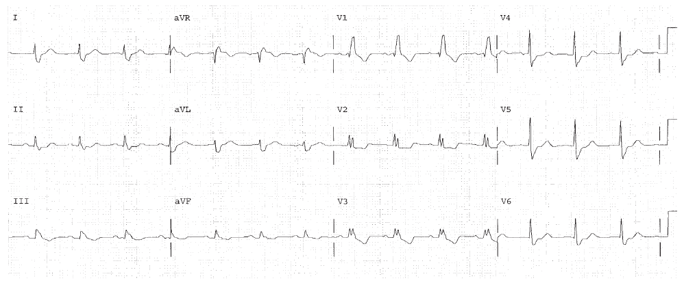
HR 83, sinus rhythm. PR interval is 0.16, normal. The QRS is 0.12 seconds indicating BBB. The end of the QRS points to the right in lead I and anteriorly in V1 indicating RBBB. The overall QRS direction is rightward since the S wave in lead I is much bigger than the R wave. This indicates LPHB.
Worksheet 2
??Examine the whole strip and determine an atrial and ventricular rate??
| Parameter | Measurement | Interpretation |
| HR | ||
| Rhythm | ||
| PR | ||
| QRS | ||
| QT | ||
| QTc | ||
| P direction | ||
| QRS direction |
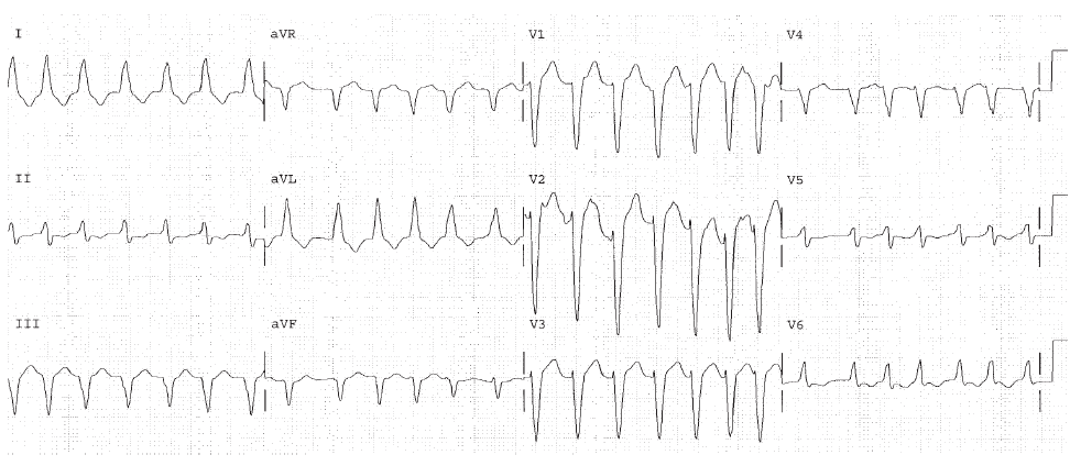
Atrial Fibrillation. Ventricular rate is 160. The PR is undefined in a fib. The QRS interval is 0.12 seconds. There is BBB. The end of the QRS is leftward (positive in lead I) and posterior (negative in lead V1) indicating LBBB. Atrial fibrillation may be associated with underlying disease such as cardiomyopathy or heart failure.
Worksheet 3
??Examine the whole strip and determine an atrial and ventricular rate??
| Parameter | Measurement | Interpretation |
| HR | ||
| Rhythm | ||
| PR | ||
| QRS | ||
| QT | ||
| QTc | ||
| P direction | ||
| QRS direction |
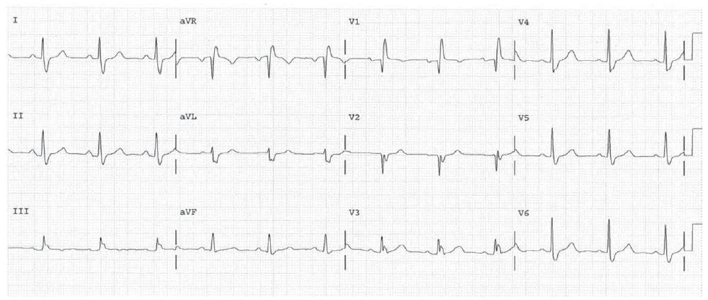
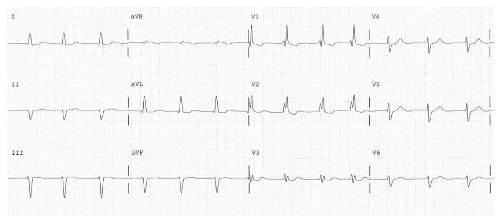
HR 68, sinus. The PR interval is 0.18, normal. The QRS is 0.14 seconds indicating BBB. The end of the QRS is negative in lead I (rightward) and positive in lead V1 (anterior) consistent with RBBB. The overall QRS points to the patients right, consistent with LPHB. The cause of RBBB and LPHB in this EKG is a septal infarction (Q waves V1 and V2), which is covered in Chapter 15.
Worksheet 4
??Examine the whole strip and determine an atrial and ventricular rate??
| Parameter | Measurement | Interpretation |
| HR | ||
| Rhythm | ||
| PR | ||
| QRS | ||
| QT | ||
| QTc | ||
| P direction | ||
| QRS direction |
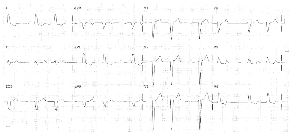
HR 72. Probable sinus rhythm with PACs and nonconducted PACs. There is not enough information on this EKG to be sure. More rhythm strips are necessary to clarify the rhythm. There is first degree AV block. The QRS interval is greater than 0.12 seconds, so BBB is present. The end of the QRS is positive in lead I and negative in V1 indicating LBBB. Clinically the rhythm needs to be proven, and the cause of LBBB determined.
Worksheet 5
??Examine the whole strip and determine an atrial and ventricular rate.??
| Parameter | Measurement | Interpretation |
| HR | ||
| Rhythm | ||
| PR | ||
| QRS | ||
| QT | ||
| QTc | ||
| P direction | ||
| QRS direction |
Atrial fibrillation. Ventricular rate 80. QRS interval is 0.11 indicating IVCD. The end of the QRS is slightly negative in lead I, and positive in lead V1. This is RIVCD (or incomplete RBBB). The overall QRS is pointing upward indicating left anterior hemiblock (LAHB).