These worksheets are for self-study only. Answers will not be evaluated.
- Complete basic measurements.
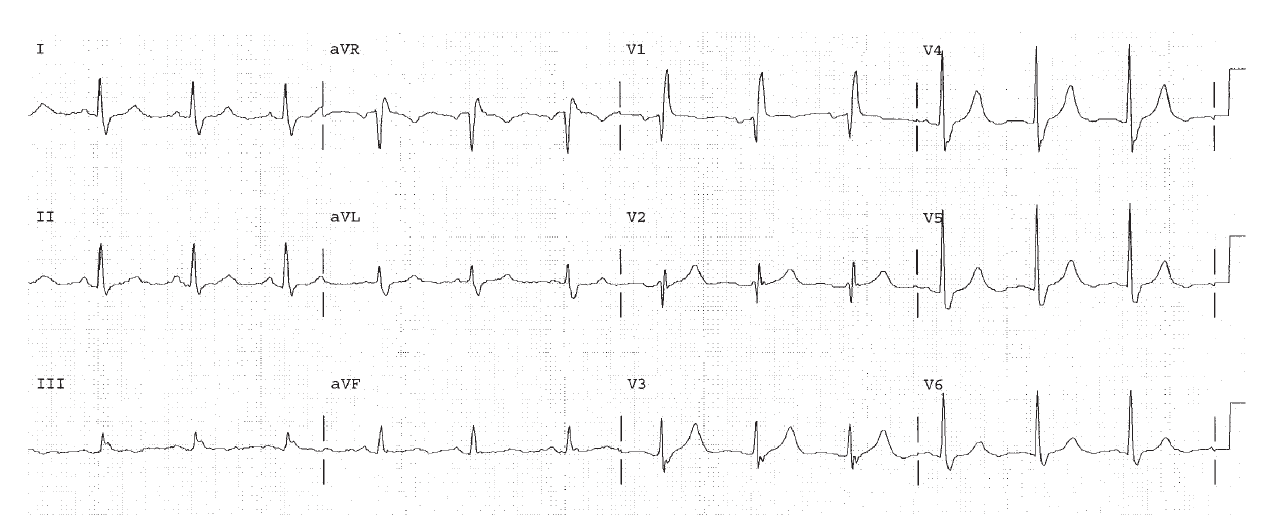
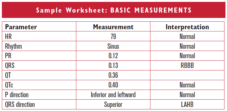
- If the QRS is ≥ 0.12 sec, diagnose BBB. Then visualize the extra piece at the end of the QRS in the frontal plane as right or left, and in the horizontal plane as anterior or posterior. Diagnose BBB further as RBBB if the end of the QRS points to the right ventricle (rightward and anteriorly). Always look for hemiblock, which may be present as well. The criteria for the diagnosis of hemiblock do not change when RBBB is present.
- Provide an interpretation.
Clinically-Based Critical Thinking: Interpretation
RBBB are LAHB are both diseases of the conduction system. The combination does not have a specific clinical correlation. The added presence of either 1°AV block (which is not present on this EKG), or symptoms of syncope would suggest the presence of further conduction disease.
Parameter | Measurement | Interpretation |
HR | ||
Rhythm | ||
PR | ||
QRS | ||
QT | ||
QTc | ||
P direction | ||
QRS direction |
Abnormal parameter | If present, note the leads or location |
Inverted T waves | |
ST depression | |
ST elevation | |
Q waves or equivalents |
HR 75, sinus. The PR is 0.16, normal. The QRS is 0.12 seconds indicating bundle branch block. The end of the QRS is negative in lead I, and so points to the right ventricle. The end of the QRS is positive in lead V1 and so points to the right ventricle. Therefore there is RBBB. The P direction is normal. The QRS overall direction is normal, so hemiblock is not present.
Worksheet 11.2
Complete the basic measurements, evaluation, and interpretation for the EKG below.
Parameter | Measurement | Interpretation |
HR | ||
Rhythm | ||
PR | ||
QRS | ||
QT | ||
QTc | ||
P direction | ||
QRS direction |
Abnormal parameter | If present, note the leads or location |
Inverted T waves | |
ST depression | |
ST elevation | |
Q waves or equivalents |
HR 65, sinus rhythm. The PR is 0.16, normal. The QRS is 0.12 seconds indicating bundle branch block. The end of the QRS is negative in lead I, pointing to the right. The end of the QRS is positive in lead V1 pointing to the right ventricle as well. This is RBBB. The P direction is normal. The overall QRS direction is rightward, since the area under the curve for the S wave is more than the R wave. This is LPHB.
Worksheet 11.3
Complete the basic measurements, evaluation, and interpretation for the EKG below.
Parameter | Measurement | Interpretation |
HR | ||
Rhythm | ||
PR | ||
QRS | ||
QT | ||
QTc | ||
P direction | ||
QRS direction |
Abnormal parameter | If present, note the leads or location |
Inverted T waves | |
ST depression | |
ST elevation | |
Q waves or equivalents |
HR 103. Sinus tachycardia. A PAC is seen in lead V1. The PR is 0.12, normal. The QRS interval is 0.13, indicating bundle branch block. The end of the QRS is negative in lead I (so pointing to the right), and positive in lead V1 (so pointing anterior). This is RBBB. The QT is 0.32. The P direction is normal. The overall QRS direction is rightward, since lead I is negative. This is LPHB. Sinus tachycardia indicates sympathetic stimulation.