


These worksheets are for self-study only. Answers will not be evaluated.
Worksheet 9.1
Calculate the atrial and ventricular rates (as able), and identify the heart rhythm.
| Atrial rate | Ventricular rate | Diagnosis |
|---|---|---|
|
|
|
|
Second degree AV block (2° AVB) Type I, Wenckebach, with four to three conduction. The PR interval gradually increases until a QRS is dropped. There are four P waves to three QRS complexes, thus a four to three ratio.
Worksheet 9.2
Calculate the atrial and ventricular rates (as able), and identify the heart rhythm.
| Atrial rate | Ventricular rate | Diagnosis |
|---|---|---|
|
|
|
|
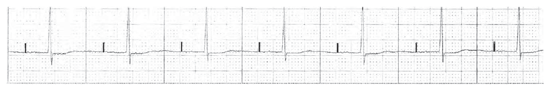
Atrial pacing. A discrete vertical spike of a pacing artifact is present 0.28 seconds before the QRS.
Worksheet 9.3
Calculate the atrial and ventricular rates (as able), and identify the heart rhythm.
| Atrial rate | Ventricular rate | Diagnosis |
|---|---|---|
|
|
|
|
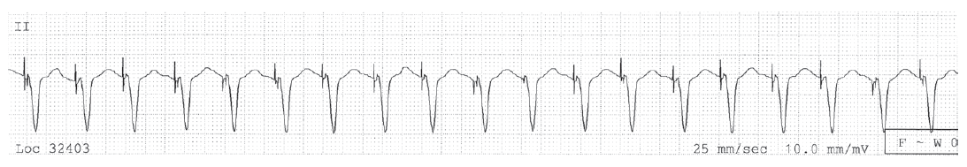
- The top strip shows ventricular pacing and a long QTc.
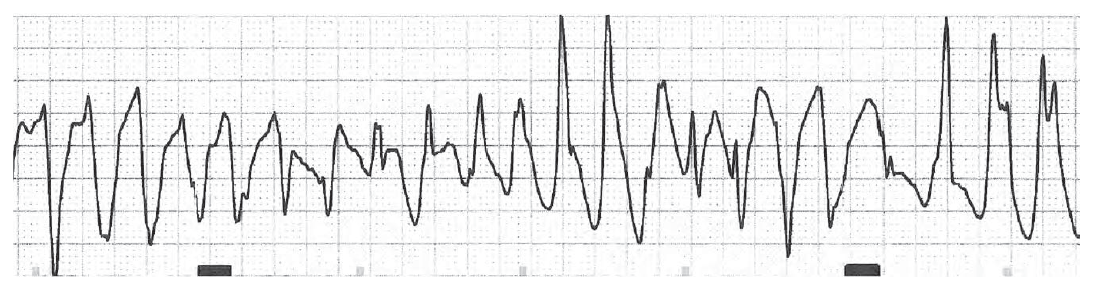
- The bottom strip taken later shows torsade.
Worksheet 9.4
Calculate the atrial and ventricular rates (as able), and identify the heart rhythm.
| Atrial rate | Ventricular rate | Diagnosis |
|---|---|---|
|
|
|
|
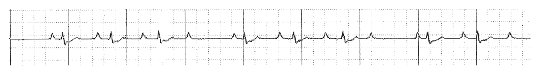
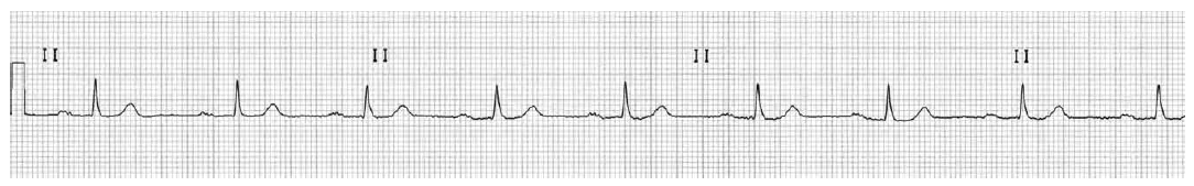
Sinus rhythm with first degree AV block.
Worksheet 9.5
Calculate the atrial and ventricular rates (as able), and identify the heart rhythm.
| Atrial rate | Ventricular rate | Diagnosis |
|---|---|---|
|
|
|
|
Atrial pacing.
Worksheet 9.6
Calculate the atrial and ventricular rates (as able), and identify the heart rhythm.
| Atrial rate | Ventricular rate | Diagnosis |
|---|---|---|
|
|
|
|
Atrial pacing followed by atrial and ventricular pacing. Note the tiny vertical pacemaker artifacts at the onset of the QRS in the last two QRS complexes.
Worksheet 9.7
Calculate the atrial and ventricular rates (as able), and identify the heart rhythm.
| Atrial rate | Ventricular rate | Diagnosis |
|---|---|---|
|
|
|
|
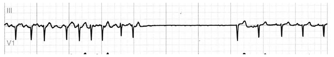
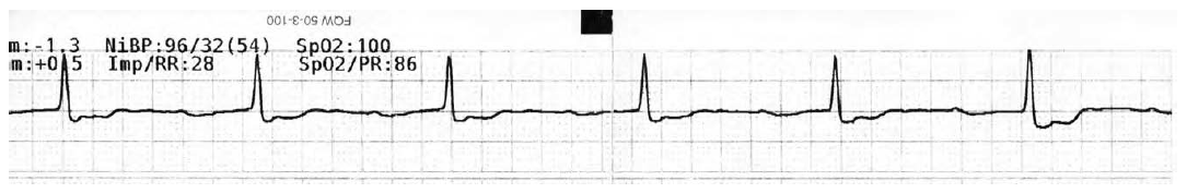
Atrial fibrillation with 4 second pause. The patient needs a pacemaker to control the pauses, and medications to control the tachycardia.
Worksheet 9.8
Calculate the atrial and ventricular rates (as able), and identify the heart rhythm.
| Atrial rate | Ventricular rate | Diagnosis |
|---|---|---|
|
|
|
|
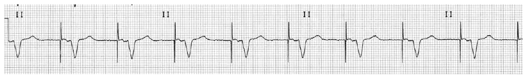
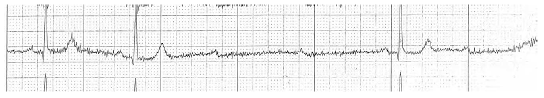
Sinus bradycardia with a very long first degree AV block. The QRS is wide suggesting bundle branch block as well. Also, there is an upward deflection hidden in the ST segment, that may represent a nonconducted P wave, which would make this second degree AV block.
Worksheet 9.9
Calculate the atrial and ventricular rates (as able), and identify the heart rhythm.
| Atrial rate | Ventricular rate | Diagnosis |
|---|---|---|
|
|
|
|
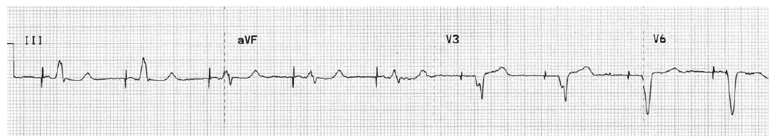
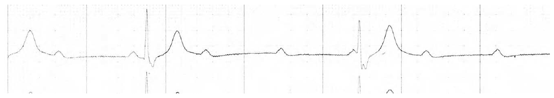
Second degree AV block, since there are P waves without QRS complexes. However there are sequential non-conducted P waves making this Type II second degree AV block, called Mobitz II, with 3:1 conduction. This requires immediate attention.
Worksheet 9.10
Calculate the atrial and ventricular rates (as able), and identify the heart rhythm.
| Atrial rate | Ventricular rate | Diagnosis |
|---|---|---|
|
|
|
|
Complete heart block with ventricular escape. The second QRS complex does not come from the preceding P wave. It is a ventricular escape beat. (Compare this to Worksheet EKG 9.9, where the QRS after the pause has a normal PR interval.)