Ventricular Arrhythmias Worksheets

Headshot of Kevin Hodges, Vice Chair, Emergency Medicine
Kevin Hodges
Vice Chair, Emergency Medicine
envelope icon phone icon
Headshot of Chris Anderson, Vice Chair, Pediatrics
Chris Anderson
Vice Chair, Pediatrics
envelope icon

These worksheets are for self-study only. Answers will not be evaluated.

Instructions for Chapter 8 Worksheets

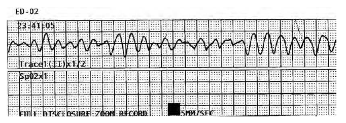
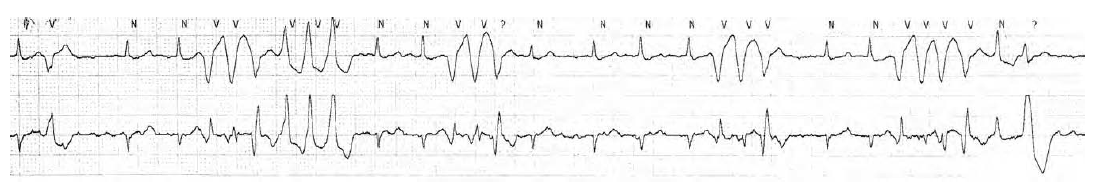
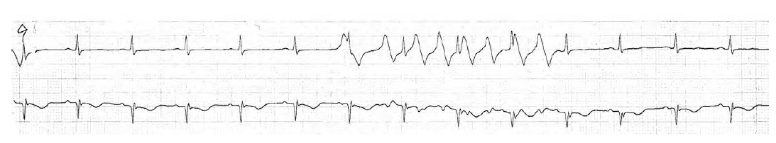
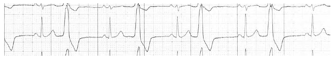
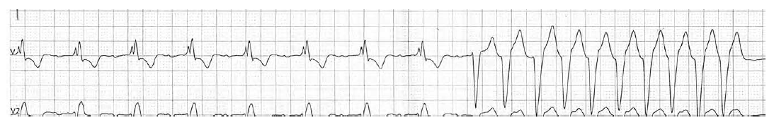
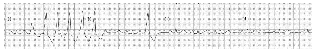
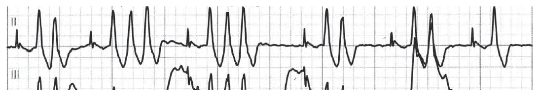
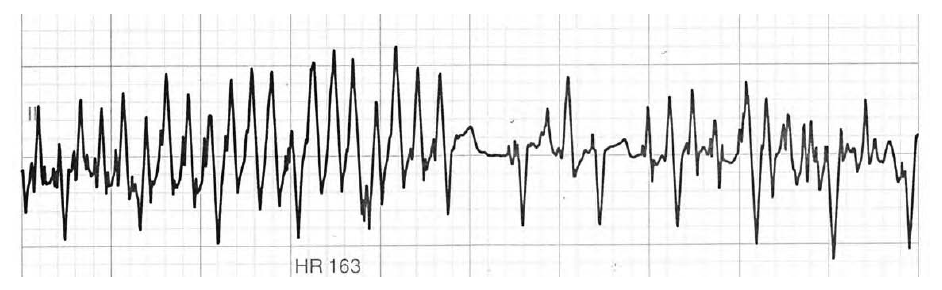
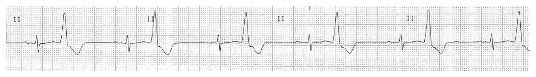
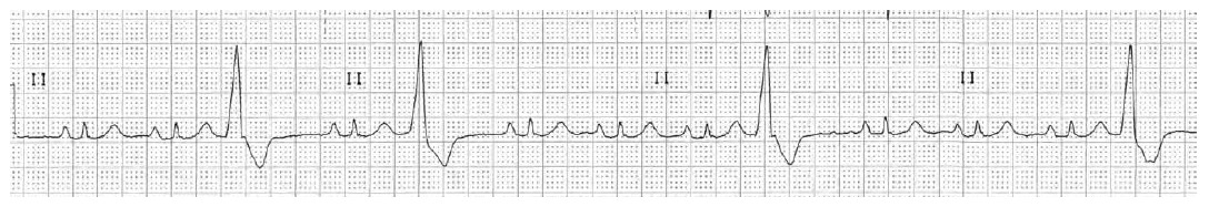
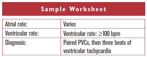
For each arrhythmia, examine the whole strip and determine an atrial and ventricular rate. The P wave samples are not long enough to confirm the underlying rhythm is sinus or to be sure of the underlying atrial rate. The ventricular rate also varies, but is more than 100 beats per minute (bpm). In addition, there is a pair of wide QRS complexes that are premature, making them paired PVCs. This is followed by a sequence of three such beats. This triplet represents ventricular tachycardia, since the QRS complexes are wide and the rate is greater than 100 bpm.


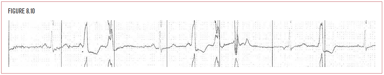
Clinical Associations:

Suspect underlying coronary disease, cardiomyopathy, or CHF. Also consider hypokalemia.

Worksheet 8.1

For each arrhythmia, examine the whole strip. Determine an atrial and ventricular rate.

Atrial rate Ventricular rate Diagnosis

Ventricular fibrillation.

Worksheet 8.2

For each arrhythmia, examine the whole strip. Determine an atrial and ventricular rate.

Atrial rate Ventricular rate Diagnosis

Polymorphous ventricular tachycardia (Torsade).

Worksheet 8.3

For each arrhythmia, examine the whole strip. Determine an atrial and ventricular rate.

Atrial rate Ventricular rate Diagnosis

Artifact. This is a dual channel recording. Both strips are taken simultaneously. Note the bottom strip shows the patient in sinus rhythm.

Worksheet 8.4

For each arrhythmia, examine the whole strip. Determine an atrial and ventricular rate.

Atrial rate Ventricular rate Diagnosis

Ventricular bigeminy.

Worksheet 8.5

For each arrhythmia, examine the whole strip. Determine an atrial and ventricular rate.

Atrial rate Ventricular rate Diagnosis

Sinus rhythm with turns into Ventricular Tachycardia. Give yourself 10 points extra if you noticed first degree AV block and Right Bundle branch block on the first part of the strip. Give yourself 20 extra points if you didn’t because you thought intervals shouldn’t be measured in Lead V1.

Worksheet 8.6

For each arrhythmia, examine the whole strip. Determine an atrial and ventricular rate.

Atrial rate Ventricular rate Diagnosis

Sinus rhythm with ventricular tachycardia, and then a PVC. The patient was asymptomatic during the recording.

Worksheet 8.7

For each arrhythmia, examine the whole strip. Determine an atrial and ventricular rate.

Atrial rate Ventricular rate Diagnosis

Atrial fibrillation with paired PVC’s and three beat runs of ventricular tachycardia.

Worksheet 8.8

For each arrhythmia, examine the whole strip. Determine an atrial and ventricular rate.

Atrial rate Ventricular rate Diagnosis

Sinus rhythm with artifact. Normal QRS complexes are seen at a regular rate at the bottom of the artifact. Of course check the patient and the EKG leads to verify this.

Worksheet 8.9

For each arrhythmia, examine the whole strip. Determine an atrial and ventricular rate.

Atrial rate Ventricular rate Diagnosis

Sinus rhythm with ventricular bigeminy.

Worksheet 8.10

For each arrhythmia, examine the whole strip. Determine an atrial and ventricular rate.

Atrial rate Ventricular rate Diagnosis

Sinus rhythm with PVC’s.