PVCs can occur in a pattern that pairs them with a normal beat. This is called ventricular bigeminy. This can occur in normal and abnormal hearts.
PVCs can occur in a pattern that pairs them with two normal beats. This is called ventricular trigeminy, or a trigeminal pattern of PVCs. This can occur in normal and abnormal hearts.
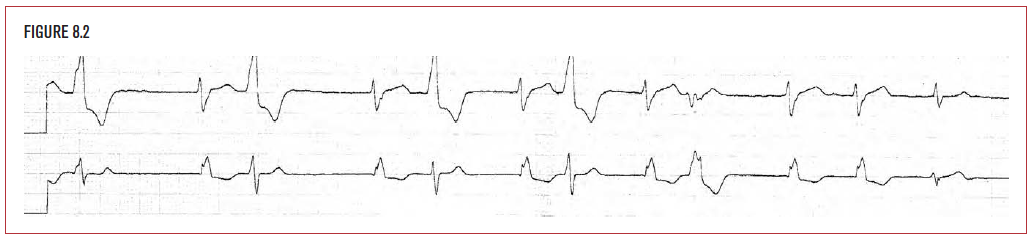
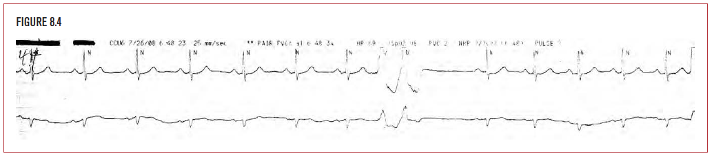
PVCs can occur in pairs, called paired PVCs or coupled PVCs. Paired PVCs have a more unstable rhythm than a single PVC and are more likely to be associated with underlying heart disease than a single PVC. Electrolyte imbalance, particularly hypokalemia, can exacerbate this.
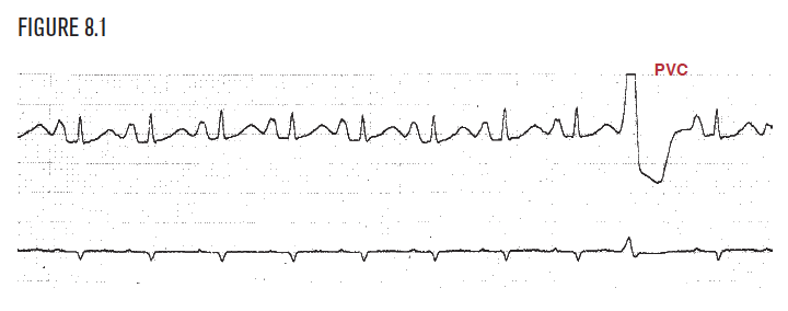
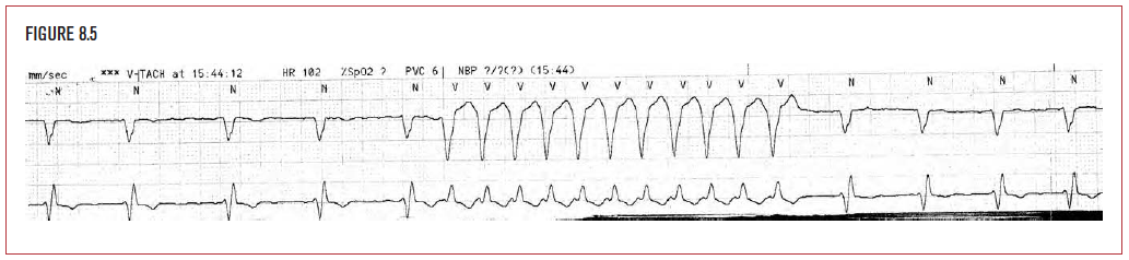
Ventricular tachycardia (VT) is a regular rhythm originating in the ventricles at a rate of greater than 100 bpm, usually 140 to 260. It has at least three beats. Figure 8.5 demonstrates a self-limited 11-beat episode of VT. VT is usually associated with underlying heart disease, such as coronary disease, cardiomyopathy, hypertension, or congestive heart failure.
Ventricular fibrillation (VF) is a fatal arrhythmia that must be rapidly terminated. It is a chaotic electrical discharge that does not effectively depolarize the ventricles and leads to death unless treated effectively and swiftly. It can be seen in acute myocardial ischemia or infarction, congestive heart failure, and cardiomyopathy. Hypokalemia increases the risk of its occurrence.
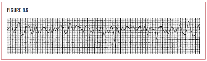
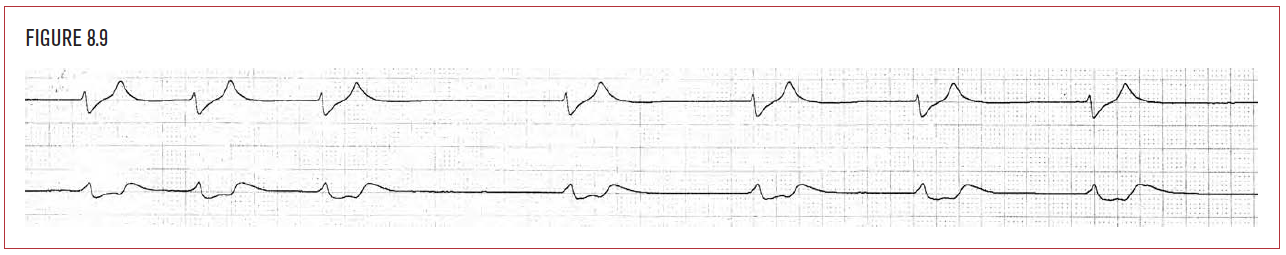
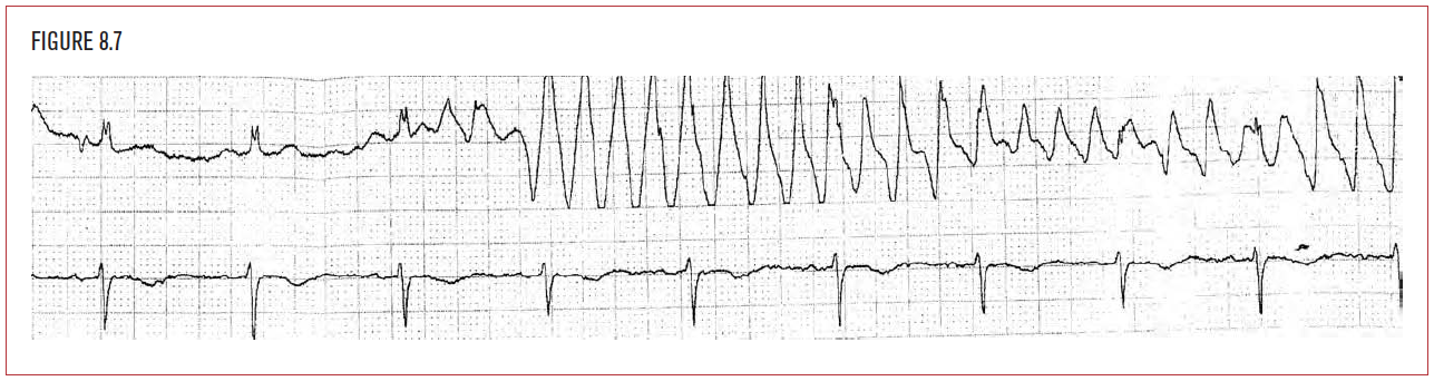
Figure 8.7 demonstrates a rhythm recorded on a two-channel recorder. Both leads are recorded at the exact same time, so any event on the top strip occurs at exactly the same time as an event exactly below it. The rhythm on top appears to be ventricular tachycardia until compared with the bottom strip, which shows sinus rhythm. Manipulation of electrodes can cause this.
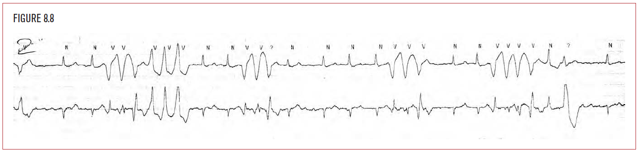
Torsade de pointe (polymorphous ventricular tachycardia) is a form of ventricular tachycardia. The diagnostic hallmark of torsade is a change in direction of the complexes. In Figure 8.8, the episode of ventricular tachycardia begins with the first three QRS complexes pointing downward and the next three pointing upward. This is associated with long QTc interval. Drugs, hypokalemia, and hypocalcemia are common causes.
Idioventricular rhythm is a slow ventricular escape rhythm characterized by wide QRS complexes, typically in a regular slow pattern with no underlying pacing in the ventricles. This is associated with underlying heart disease and can be worsened by beta-blockers or calcium channel blockers.