


These worksheets are for self-study only. Answers will not be evaluated.
Instructions for Chapter 7 Worksheets
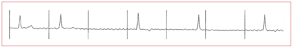
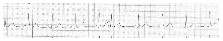
For each arrhythmia, examine the whole strip. Determine an atrial and ventricular rate. In the figure below, the atrial rate in indeterminate. The ventricular rate is irregularly irregular and consistent with atrial fibrillation. The correct way to calculate the heart rate in atrial fibrillation is to count the number of QRS complexes in a random six second sample and then multiply that number by ten to get the ventricular rate in beats per minute (bpm). The figure below shows 4 QRS complexes in a six-second interval. Four multiplied by 10 would yield a ventricular rate of 40 bpm.

Clinical Associations:
Hyperthyroidism, pulmonary embolism, COPD, hypertension, valvular heart disease, CHF.
Worksheet 7.1
For each arrhythmia, examine the whole strip. Determine an atrial and ventricular rate.
| Atrial rate | Ventricular rate | Diagnosis |
|---|---|---|
|
|
|
|
Atrial flutter. Three second pause demonstrates atrial flutter waves, but no QRS complexes.
Worksheet 7.2
For each arrhythmia, examine the whole strip. Determine an atrial and ventricular rate.
| Atrial rate | Ventricular rate | Diagnosis |
|---|---|---|
|
|
|
|
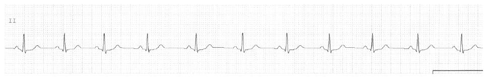
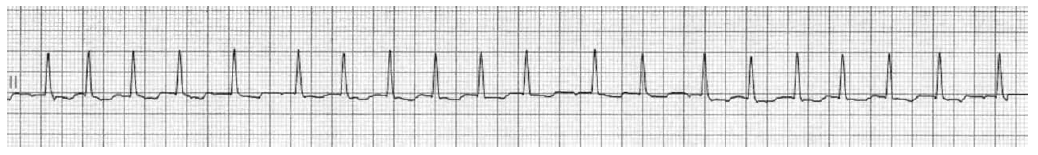
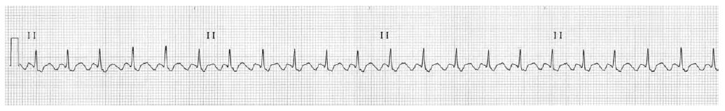
Sinus arrhythmia. Rate varies from 60 to 75.
Worksheet 7.3
For each arrhythmia, examine the whole strip. Determine an atrial and ventricular rate.
| Atrial rate | Ventricular rate | Diagnosis |
|---|---|---|
|
|
|
|
Atrial fibrillation. Ventricular rate 180.
Worksheet 7.4
For each arrhythmia, examine the whole strip. Determine an atrial and ventricular rate.
| Atrial rate | Ventricular rate | Diagnosis |
|---|---|---|
|
|
|
|
Sinus rhythm with a premature atrial contraction PAC. Examine the T wave before the premature QRS. This T wave is deformed because a hidden P wave is present.
Worksheet 7.5
For each arrhythmia, examine the whole strip. Determine an atrial and ventricular rate.
| Atrial rate | Ventricular rate | Diagnosis |
|---|---|---|
|
|
|
|
Sinus rhythm turns into rapid atrial fibrillation.
Worksheet 7.6
For each arrhythmia, examine the whole strip. Determine an atrial and ventricular rate.
| Atrial rate | Ventricular rate | Diagnosis |
|---|---|---|
|
|
|
|
Atrial flutter with varying conduction (2:1 and 4:1).
Worksheet 7.7
For each arrhythmia, examine the whole strip. Determine an atrial and ventricular rate.
| Atrial rate | Ventricular rate | Diagnosis |
|---|---|---|
|
|
|
|
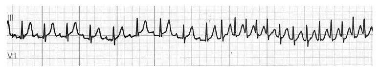
Narrow QRS tachycardia, irregular, probably atrial fibrillation. Always get long strips off telemetry to double check.
Worksheet 7.8
For each arrhythmia, examine the whole strip. Determine an atrial and ventricular rate.
| Atrial rate | Ventricular rate | Diagnosis |
|---|---|---|
|
|
|
|
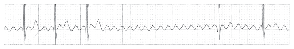
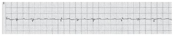
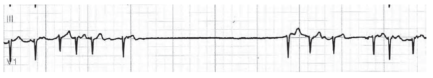
Atrial fibrillation with a pause of greater than 4 seconds. This patient has fast and low heart rates. A pacemaker will be needed to control the low heart rate, and antiarrhythmic drugs such as digitalis, beta blockers, and calcium channel blockers may be needed to control the fast heart rates.
Worksheet 7.9
For each arrhythmia, examine the whole strip. Determine an atrial and ventricular rate.
| Atrial rate | Ventricular rate | Diagnosis |
|---|---|---|
|
|
|
|
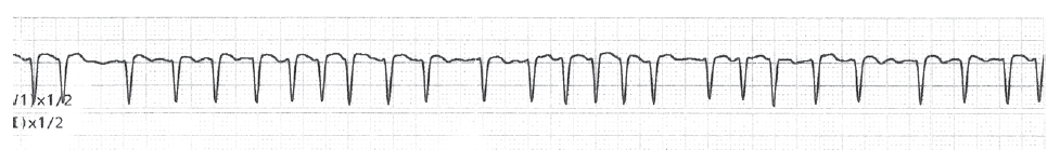
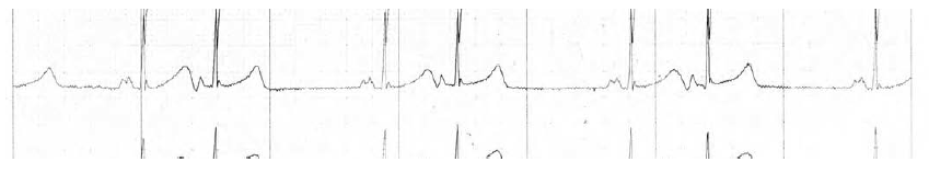
Atrial flutter with 2:1 block. Erase the QRS complexes from your mind, and you can see the sawtooth pattern of atrial flutter.
Worksheet 7.10
For each arrhythmia, examine the whole strip. Determine an atrial and ventricular rate.
| Atrial rate | Ventricular rate | Diagnosis |
|---|---|---|
|
|
|
|
Atrial bigeminy. There may be a nonconducted PAC hidden in the second T wave of each pair, accounting for the pauses.