Gluteal region, posterior thigh, and popliteal fossa
Pertinent osteology Please review the anatomy of the acetabulum, posterior hip bone, sciatic notches, and femur. Figure 24.1 Osteology of the os coxae. Figure 24.2 Sciatic foramina. Figure 24.3 Osteology of the femur. Gluteal region Physically, the gluteal region is part of the trunk, but functionally, it is clearly part of the limb. The gluteal […]
The hip joint
Figure 23.1 The hip joint is the articulation between the round femoral head and the concave acetabulum (“little vinegar cup”). The lunate surface is the articular surface of the acetabulum, forming an arc that fills ¾ of the acetabular cup. It is covered with articular cartilage. The acetabulum is deepened by the acetabular labrum, a […]
Anterior and medial compartments of the thigh
Optional Reading Clinically Oriented Anatomy, 8th ed., Anterior and medial regions of thigh section through Surface anatomy of anterior and medial regions of thigh. Compartmentalization of the thigh The deep fascia, intermuscular septa, and femur together define anterior and posterior compartments in the thigh. The anterior compartment contains muscles that flex the hip and extend […]
Pelvic vessels, nerves, and lymphatics
Optional Reading Clinically Oriented Anatomy, 8th ed., chapter 6, Neurovascular structures of pelvis section through Clinical box: Neurovascular structures of pelvis. Vascular supply of the pelvis Arteries The internal iliac arteries are the prime sources of blood to pelvic structures. They also supply musculoskeletal structures outside the pelvic cavity (hip and gluteal regions). Arising from […]
Pelvic viscera
Optional Reading Clinically Oriented Anatomy, 8th ed., Pelvic viscera section through Lymphatic drainage from female pelvic viscera. The “lay of the land”: The pelvic viscera are below the peritoneum, surrounded by visceral pelvic fascia, and separated from one another by subperitoneal pelvic connective tissue “packing material.” Most of the organs in the pelvic cavity (rectum, […]
The pelvic cavity and pelvic skeleton
Optional Reading Clinically Oriented Anatomy, 8th ed., chapter 6, Introduction to pelvis and perineum section through The bottom line: Pelvic cavity, pelvic peritoneum, and pelvic fascia. The pelvis is the region of transition where the trunk and lower limbs meet. Since the word pelvis comes from the Latin term for basin, it is used more […]
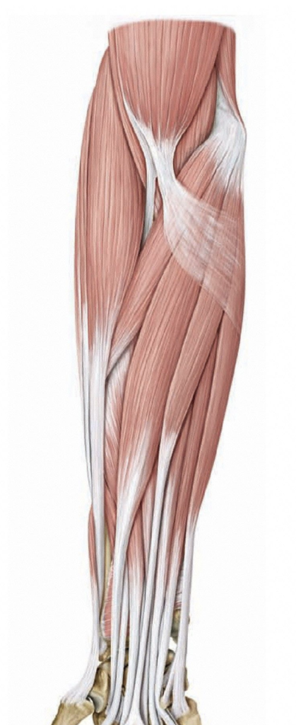
Posterior forearm
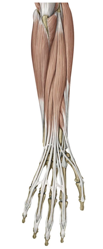
Cubital fossa and anterior forearm
Autonomic innervation of thoracic organs
Now let’s take the principles of the ANS that we learned in Principles of the autonomic nervous system (ANS) and apply them specifically to innervating thoracic organs. Autonomic nerve plexuses Autonomic motor and visceral afferent fibers reach and leave thoracic organs through autonomic nerve plexuses. Concept In autonomic plexuses, parasympathetic and sympathetic fibers and visceral […]
Heart and pericardium
Location of the heart and pericardium The heart and pericardium are located in the mediastinum, resting atop the diaphragm, between the lungs and pleural cavities. Owing to its development, most of the heart is located to the left side of the body’s midline. The size of the heart is described as that of the person’s […]