Protected: Lab 31: Dissection: Infratemporal Fossa and Floor of Mouth; Bony Anatomy of Pterygopalatine Fossa
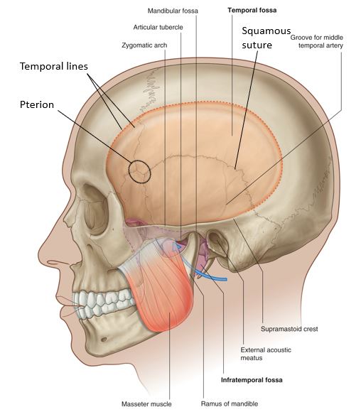
There is no excerpt because this is a protected post.
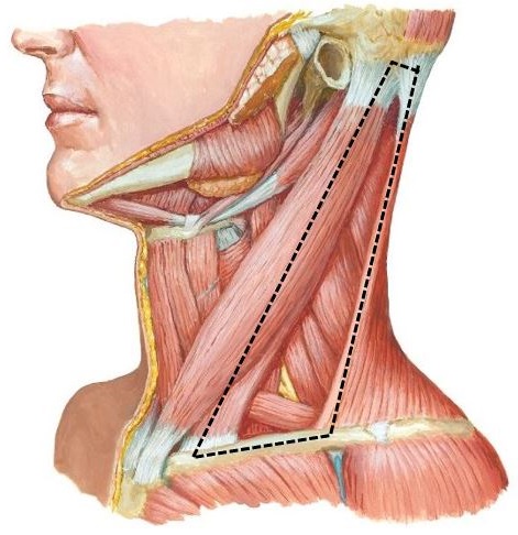
Protected: Lab 29: Neck: Anterior Triangle of Neck
There is no excerpt because this is a protected post.
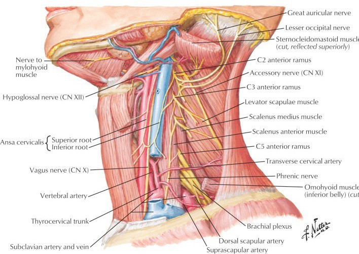
Protected: Lab 30: Dissection: Posterior Triangle of Neck, Deep Neck, and Root of Neck
There is no excerpt because this is a protected post.
Protected: Lab 28: Dissection: Orbit and Ear
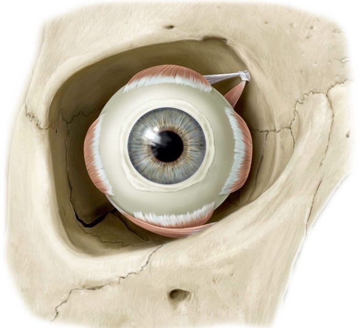
There is no excerpt because this is a protected post.
Protected: Lab 27: Face, Parotid Gland, and Superficial Neck
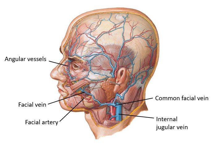
There is no excerpt because this is a protected post.
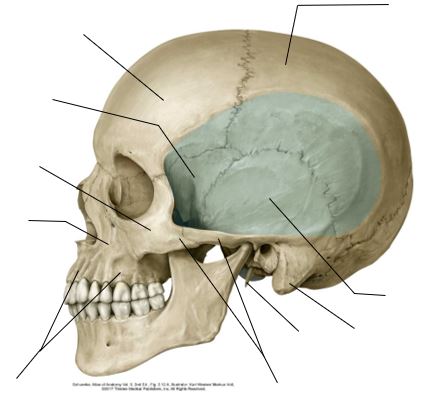
Protected: Lab 26: Scalp, Cranial Cavity, and Meninges
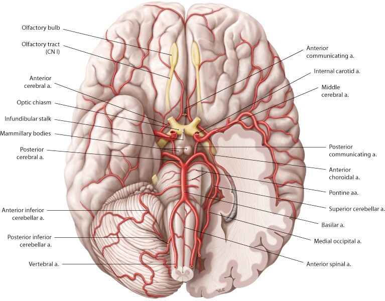
There is no excerpt because this is a protected post.
Protected: Lab 24: Peritoneal Cavity and Supracolic Region
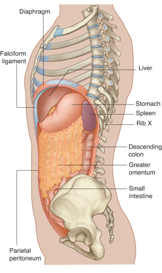
There is no excerpt because this is a protected post.
Protected: Lab 25: Infracolic Region
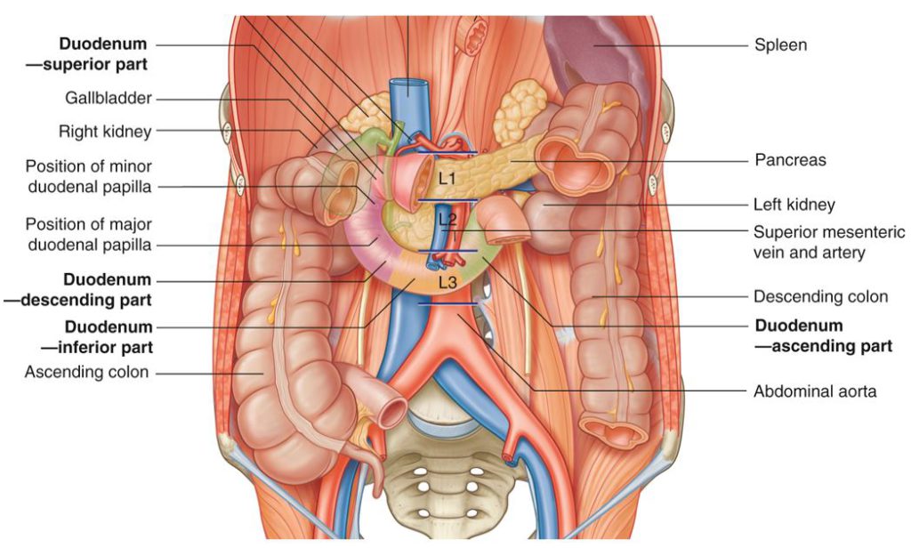
There is no excerpt because this is a protected post.
Protected: Lab 23, Station 5: Oral Region and Salivary Glands—Lateral View
There is no excerpt because this is a protected post.
Protected: Lab 23, Station 4: Oral Cavity and Pharynx—Sagittal View
There is no excerpt because this is a protected post.